

Depth learning-based abnormal chest radiograph intelligent identification method and system
An intelligent recognition and deep learning technology, applied in the field of image recognition, can solve problems such as time-consuming, dependence on personal experience and working time, and low efficiency
- Summary
- Abstract
- Description
- Claims
- Application Information
AI Technical Summary
Problems solved by technology
Method used
Image

Examples
Embodiment Construction
[0025] In order to make the technical means, creative features, goals and effects achieved by the present invention easy to understand, the present invention will be further described below in conjunction with specific illustrations.
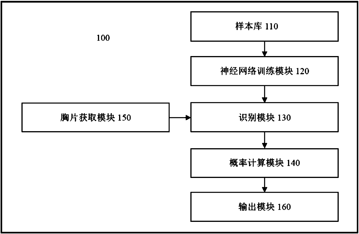
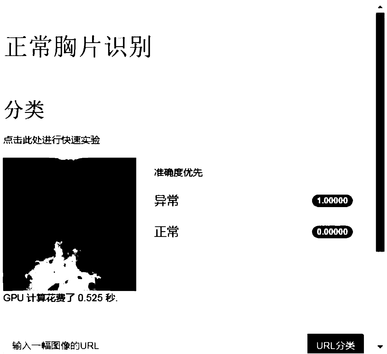
[0026] Aiming at the identification of abnormal features in chest radiographs, this example program performs automatic identification to improve efficiency and effectively avoid unrecognized phenomena; on this basis, the deep learning method is used to independently learn the abnormal image features of chest radiographs to achieve accurate identification , effectively improving the recognition accuracy.
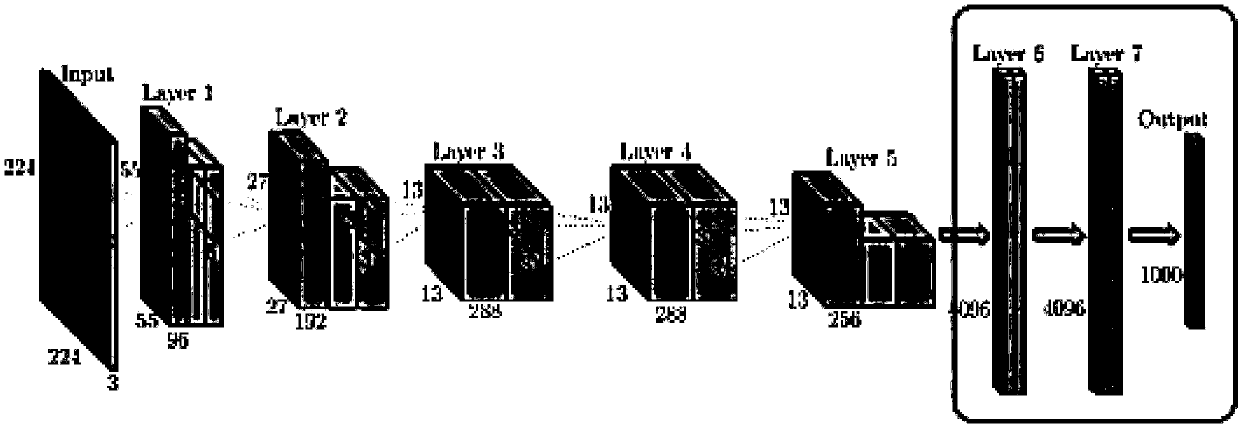
[0027] Specifically, this program uses a large number of manually labeled samples to train a deep neural network, so that it can learn the abnormal image features of chest radiographs autonomously, so as to identify the abnormal image features in chest radiographs.
[0028] The large number of samples used here contains positive samples of abno...
PUM
 Abstract
Description
Claims
Application Information