Dynamic constraint graph search-based algorithm for acquiring physiological parameters in retina OCT image

A technology of dynamic constraints and physiological parameters, applied in image data processing, image enhancement, image analysis, etc., can solve the problems of inaccuracy, waste of time, algorithm robustness and accuracy, and achieve accurate results and accurate measurements. robust effect

Active Publication Date: 2018-03-27
SUZHOU BIGVISION MEDICAL TECH CO LTD
View PDF4 Cites 3 Cited by
  • Summary
  • Abstract
  • Description
  • Claims
  • Application Information

AI Technical Summary

Problems solved by technology

[0002] At present, the existing retinal image analysis methods centered on the optic nerve head, especially the acquisition methods of some physiological parameters of the retina mainly rely on manual measurement, which is a waste of time, and due to differences in the cognitive level of the measurement people, the retinal Acquisition of a series of physiological parameters is slow and imprecise
At the same time, the few existing retinal image analysis and physiological parameter measurement algorithms at home and abroad can only obtain part of the parameter information, and the robustness and accuracy of the algorithm are not ideal

Method used


Image

  • Dynamic constraint graph search-based algorithm for acquiring physiological parameters in retina OCT image
  • Dynamic constraint graph search-based algorithm for acquiring physiological parameters in retina OCT image
  • Dynamic constraint graph search-based algorithm for acquiring physiological parameters in retina OCT image

Examples


Embodiment 1

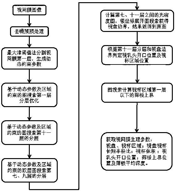
[0031] figure 2 It is a retinal image and structure centered on the optic head. The optic head is located about 3 mm in the temporal side of the macular area of ​​the retina, with a diameter of about 1.5 mm. The visual fibers on the retina converge here and pass through to the optic center. There is a small depression in the center that becomes the optic cup. The optic head is the starting end of the optic nerve fibers that aggregate to form the optic nerve. There are no visual cells and therefore no vision. It is a physiological blind spot in the visual field. The reticular structure that optic nerve fibers pass through in the process of leading to the visual center is the cribriform plate. figure 1 The middle left image is a slice of the retinal image centered on the optic nerve head, and the right side is an annotated image of each physiological region, wherein the upper layer of the depression in the middle is the layered surface of the first layer of the retina, and the...


PUM


Abstract

The invention discloses a dynamic constraint graph search-based algorithm for acquiring physiological parameters in a retinal OCT image. The algorithm is characterized in that the algorithm comprisesthe following steps of: S01, image preprocessing: a three-dimensional retina OCT image with an optic nerve head adopted a center is subjected to filtering and de-noising processing; S02, retina layering: the first layer of a retina is roughly separated out, dynamic constraint parameters are obtained, the first layer of the retina is accurately separated out on the basis of the dynamic constraint parameters, alignment processing is performed on the three-dimensional image, the eleventh layer of the retina is accurately separated out, and the seventh layer and ninth layer of the retina are accurately separated out; S03, optic disc and optic cup region segmentation; S04, cribriform plate upper boundary segmentation; and S05, the acquisition of physiological parameter information in the retinaimage. With the algorithm of the present invention adopted, the retina OCT image with the optic nerve head adopted the center can be accurately segmented, and relevant physiological parameters can beobtained.

Description

technical field [0001] The invention relates to an algorithm for obtaining physiological parameters in retinal OCT images based on dynamic constraint graph search, and belongs to the technical field of image processing and analysis. Background technique [0002] At present, the existing retinal image analysis methods centered on the optic head, especially the acquisition methods of some physiological parameters of the retina, mainly rely on manual manual measurement, which is time-consuming, and due to the differences in the cognitive level of the measurement people, the retinal A series of physiological parameters are acquired slowly and imprecisely. At the same time, the few existing retinal image analysis and physiological parameter measurement algorithms at home and abroad can only obtain partial parameter information, and the robustness and accuracy of the algorithms are not ideal. SUMMARY OF THE INVENTION [0003] The technical problem to be solved by the present in...

Claims


Application Information

Patent Timeline
Patent Type & Authority Applications(China)
IPC IPC(8): G06T7/00G06T7/12G06T7/162
CPCG06T7/0012G06T7/12G06T7/162G06T2207/10101G06T2207/20016G06T2207/20072G06T2207/30041
Inventor 陈新建张秀兰俞凯李飞
Owner SUZHOU BIGVISION MEDICAL TECH CO LTD
Why Eureka
Try Eureka