Method for preparing 3D printed model of human airway and cervical vertebra

A 3D printing and cervical spine technology, applied in the field of 3D printing, can solve problems such as inability to understand the structure and complex airway structure, and achieve the effects of reducing the discomfort of intubation, facilitating clinical teaching, and improving the success rate of intubation

Active Publication Date: 2018-06-01
PEKING UNIV THIRD HOSPITAL
View PDF4 Cites 0 Cited by
  • Summary
  • Abstract
  • Description
  • Claims
  • Application Information

AI Technical Summary

Benefits of technology

This invention relates to methods that help train medical professionals better manage complex breathing problems during surgeries or procedures while reducing patient complications such as difficulty breathe due to congenital heart disease (CWD). These techniques involve restoring complete respiratory functions through various means like artificial lungs or prostheses made from synthetic material. Additionally, it suggests adding colorings to improve visual recognition when navigating complicated areas around them. Overall, these technical improvements aimed at improving lung function are useful tools for training healthcare providers to treat more challenging cases effectively without causing pain or injury.

Problems solved by technology

This patented technical problem addressed in this patents relates to improving methods for safely inserting medical devices into narrow spaces within the body without causing harm or injury from accidentally damaging other structures around them (such as blood vessels).

Method used


Image

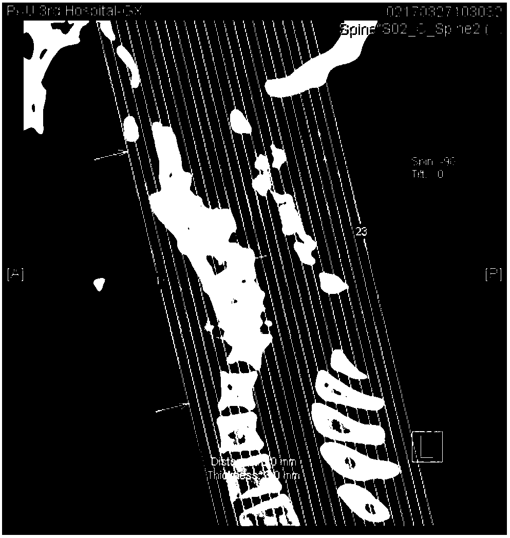
  • Method for preparing 3D printed model of human airway and cervical vertebra
  • Method for preparing 3D printed model of human airway and cervical vertebra
  • Method for preparing 3D printed model of human airway and cervical vertebra

Examples


Embodiment Construction

[0035] The present invention will be further described in detail below in conjunction with the accompanying drawings and specific embodiments. There is no software innovation in the present invention.

[0036] The invention provides a method for preparing a human airway and cervical vertebra 3D printing model, comprising the following steps:

[0037] Step 1. Following the principles of medical ethics, obtain the images and data obtained from the original CT tomography of the patient's intended printing site;

[0038] The image is in Dicom format or JPG format, and the tomogram format and image size must be uniform;

[0039] CT is medical data information, and what 3D printing needs is a closed solid model, so the image is converted through Mimics software.

[0040] Step 2. Carry out three-dimensional reconstruction according to the obtained image and data of the part to be printed;

[0041] What 3D printing needs is a closed solid model, so we need to convert the scanned im...


PUM


Abstract

The invention provides a method for preparing a 3D printed model of a human airway and cervical vertebra. The method comprises the following steps: subjecting a 3D printed hollow airway to 3D solid scanning so as to obtain a 3D scanned image and 3D image data, comparing the obtained 3D scanned image and 3D image data with original CT scanning data, and evaluating the accuracy of the printed hollowairway; and then assembling a printed cervical vertebra and adjacent appendicular bony tissue thereof with the hollow airway. The method provided by the invention can completely reproduce the airway,the cervical vertebra and adjacent anatomical structures, facilitates preoperative simulation of endotracheal intubation in patients with difficult airways, and formulates detailed airway managementstrategies; a hard material is used for printing to print bony structures, and a soft material is used for printing the airway, so the body structure of human beings is authentically reproduced; and to highlight the airway, the airway printing material can be of different colors to facilitate clinical teaching.

Description


Claims


Application Information

Patent Timeline
Owner PEKING UNIV THIRD HOSPITAL
Why Eureka
Try Eureka