Pseudorabies virus GE gene major antigen-epitope region recombinant protein preparation and colloidal-gold immunochromatographic strip

A main antigenic epitope, pseudorabies virus technology, applied in the direction of virus/phage, virus, virus peptide, etc., to achieve the effect of strong specificity, low cost and wide application

Inactive Publication Date: 2019-12-06
SHANGHAI VETERINARY RES INST CHINESE ACAD OF AGRI SCI +1
View PDF0 Cites 3 Cited by
  • Summary
  • Abstract
  • Description
  • Claims
  • Application Information

AI Technical Summary

Problems solved by technology

[0004] The neutralization test is generally considered the most standard pseudorabies detection method in the academic circles, but in the actual detection work, the neutralization te

Method used


Image

  • Pseudorabies virus GE gene major antigen-epitope region recombinant protein preparation and colloidal-gold immunochromatographic strip
  • Pseudorabies virus GE gene major antigen-epitope region recombinant protein preparation and colloidal-gold immunochromatographic strip
  • Pseudorabies virus GE gene major antigen-epitope region recombinant protein preparation and colloidal-gold immunochromatographic strip

Examples


Embodiment 2

[0027] Example 2 Preparation and purification of protein

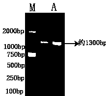
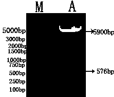
[0028] (1) Expression of recombinant plasmids

[0029] (1) Inoculate the host strain BL21(DE3) containing the recombinant plasmid pET-32a(+)-GE with correct sequencing into 250 mL of LB liquid medium containing 50 μg / mL Amp+, and place it in a 37°C incubator at 250 Shake culture at r / min until the logarithmic phase of bacterial growth (OD600 is about 0.6).

[0030] (2) Add IPTG solution with a final concentration of 1 mmol / L and induce for 8 h. Before induction, 1 h, 2 h, 3 h, 4 h, 6 h, and 8 h after induction, respectively, 1 mL of bacterial solution was taken, Carry out SDS-PAGE electrophoresis identification, and observe the best induced expression time.

[0031] (3) After 4 hours of induction, the bacterial solution was centrifuged at 12 000 r / min for 15 minutes at 4 °C to collect bacterial precipitates.

[0032] (4) The bacterial pellet was resuspended in 12 mL 1×PBS, and after repeated freezing and thawing thr...

Embodiment 3

[0056] Immunogenicity identification of embodiment 3 recombinant protein

[0057] To test the immunogenicity of recombinant proteins, follow the steps below:

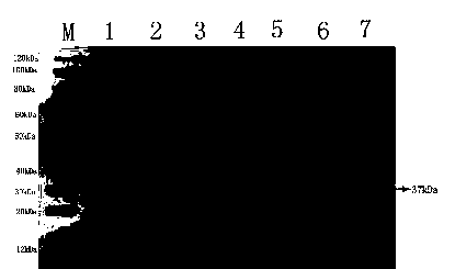
[0058] (1) Perform SDS-PAGE electrophoresis on the recombinant protein after purification and dialysis. After electrophoresis, transfer the recombinant protein to a nitrocellulose membrane with a current of 260 mA for 1 h under ice bath conditions;

[0059] (2) Sealing of nitrocellulose membrane: Weigh 5 g of skimmed milk powder and dissolve it in 100 mL of 1×PBST, soak the nitrocellulose membrane in the solution, and seal at 37 °C for 2 h;

[0060] (3) Primary antibody incubation: After blocking, wash off unbound skim milk powder with PBST (10 min each time, 4 times), then use PRV Ab(+) (1:100 dilution) as the primary antibody, and incubate at 37 °C 2 hours;

[0061] (4) Secondary antibody incubation: wash off the unbound primary antibody with PBST (10 min each time, 4 times), use goat anti-pig IgG (1:3000 dilution) ...

Embodiment 4

[0063] The preparation of embodiment 4 colloidal gold immunochromatography test strips

[0064] 4.1 Protein expression Purified recombinant protein was prepared as in Example 2

[0065] 4.2 Spotting on nitrocellulose membrane: Dilute recombinant GE protein to 0.5mg / ml for streaking of detection line (T line), dilute mouse anti-His tag monoclonal antibody to 0.5mg / ml for quality The underline of the control line (C line). Stick the NC film on the bottom plate, and then use the colloidal gold spotting system to draw the line. The amount of the line is 1µl / cm. Desiccant, valid for 15 months.

[0066] 4.3 Firing of colloidal gold

[0067] Burn colloidal gold according to the sodium citrate reduction method: soak all glassware used for firing and preserving colloidal gold in an acid tank for 24 hours, take them out, rinse them and dry them for later use. Take 100mL of deionized water and 1mL of 1% chloroauric acid, and heat to boiling. Add 1.5 mL of 1% trisodium citrate dropwi...


PUM


Abstract

The invention provides a pseudorabies virus GE protein and preparation of a colloidal-gold immunochromatographic strip thereof. The pseudorabies virus colloidal-gold immunochromatographic strip is provided with a carrier plate, a sample pad, a colloidal-gold conjugate pad, a laminate membrane, a detection line, a quality-control line and an absorption pad. The expressed recombinant protein existedin a supernatant is easy to purify. The detection method established by taking the recombinant protein, which is purified by affinity chromatography, as a diagnostic antigen is low in cost and simpleand convenient, and especially the diagnostic antigen concentrates major antigen-epitopes of the gE antigen. The detection sensitivity and specificity of the recombinant protein are both higher thanthe intact gE protein.

Description

technical field [0001] The present invention relates to the technical field of animal virology and immunology detection, more specifically to the construction of a viral gE protein used to detect pseudorabies antibodies in pig serum, and also to a method for preparing immune colloidal gold test paper using the above-mentioned protein, It also relates to the use of an immune colloidal gold test paper for detecting pseudorabies virus gE protein in pig serum. Background technique [0002] Pseudorabies Virus (PRV) is a herpes virus that causes fever, itching (except pigs) and acute encephalomyelitis in various livestock and wild animals such as cattle, sheep, pigs, dogs and cats. The symptoms of the disease caused by the virus resemble rabies, so it is called pseudorabies. Pigs are the storage host and source of infection of PRV. Healthy pigs can be infected with the disease through direct contact with diseased or infected pigs. It mainly causes sow reproductive disorders, abo...

Claims


Application Information

Patent Timeline
IPC IPC(8): C07K14/03C12N15/38G01N33/569G01N33/532G01N33/558G01N33/58
CPCC07K14/005C12N2710/16722G01N33/532G01N33/558G01N33/56983G01N33/587G01N2333/032
Inventor 朱传刚周志平冒丽刘冀吴思敏纪荣毅沈元曦
Owner SHANGHAI VETERINARY RES INST CHINESE ACAD OF AGRI SCI
Why Eureka
Try Eureka