Endoscopic OCT-Raman dual-mode imaging device and imaging method

A dual-mode imaging and imaging technology, applied in the field of endoscopy, can solve the problems of low visualization, affecting the intensity of Raman signals, and uniform image distortion. Effect

Active Publication Date: 2020-12-18
NANJING UNIV OF AERONAUTICS & ASTRONAUTICS
View PDF19 Cites 6 Cited by
  • Summary
  • Abstract
  • Description
  • Claims
  • Application Information

AI Technical Summary

Problems solved by technology

[0004] The existing OCT-Raman endoscopic probe has the following problems: First, although the existing Raman endoscopic system can obtain the Raman spectrum of the endoscopic image and its corresponding position, the Raman spectrum itself exists in the form of spectral lines , complex data analysis is required to realize the analysis of lesions, the degree of correlation with the imaging endoscopic image is low, the position correspondence is not obvious, the degree of visualization is low, and the abnormality and lesion cannot be clearly judged
Secondly, in the cavity endoscopy system, the imaging position of the endoscop

Method used


Image

  • Endoscopic OCT-Raman dual-mode imaging device and imaging method
  • Endoscopic OCT-Raman dual-mode imaging device and imaging method
  • Endoscopic OCT-Raman dual-mode imaging device and imaging method

Examples


Embodiment Construction

[0034] In order to make the object, technical solution and advantages of the present invention clearer, the present invention will be further described in detail below in conjunction with the accompanying drawings and embodiments. It should be understood that the specific embodiments described here are only used to explain the present invention, not to limit the present invention.

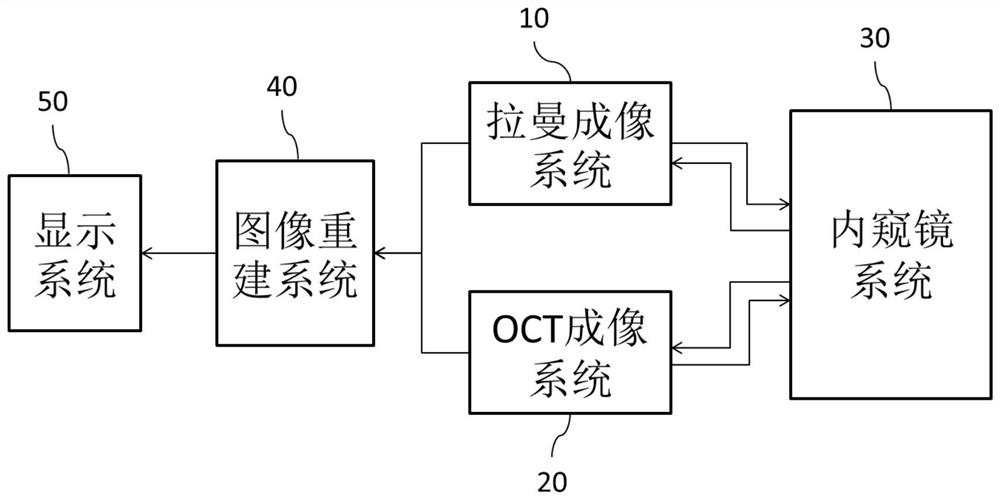
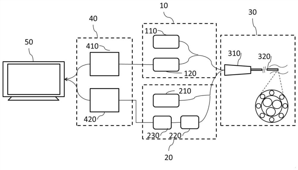
[0035] figure 1 It is a schematic diagram of the overall structure of the endoscopic OCT-Raman dual-mode imaging device of the embodiment of the present invention, figure 2It is a connection schematic diagram of the endoscopic OCT-Raman dual-mode imaging device of the embodiment of the present invention. In this embodiment, the endoscopic OCT-Raman dual-mode imaging device includes a Raman imaging system 10 , an OCT imaging system 20 , an endoscope system 30 , an image reconstruction system 40 and a display system 50 . Endoscope system 30 is connected with Raman imaging system 10 and OCT imaging...


PUM


Abstract

The invention discloses an endoscopic OCT-Raman dual-mode imaging device and an imaging method. The endoscopic OCT-Raman dual-mode imaging device comprises a Raman imaging system, an OCT imaging system, an endoscope system, an image reconstruction system and a display system, wherein the endoscope system is connected with the Raman imaging system and the OCT imaging system respectively, the Ramanimaging system and the OCT imaging system are both connected with the image reconstruction system, and the image reconstruction system is connected with the display system. According to the OCT-Ramandual-mode imaging device disclosed by the invention, a three-dimensional image with high optical resolution can be obtained, the spectral line form existence mode of a Raman spectrum is changed, the Raman spectrum is associated with OCT three-dimensional imaging, reconstruction of an OCT cavity tiled three-dimensional image and the corresponding Raman three-dimensional image is realized, lesion position correspondence is more obvious, the imaging visualization degree is improved, and accurate and rapid acquisition of OCT imaging data and Raman signals is realized.

Description

technical field [0001] The invention belongs to the technical field of endoscopes, in particular to an endoscope OCT-Raman dual-mode imaging device and an imaging method. Background technique [0002] As a non-invasive or minimally invasive imaging method, endoscopy can penetrate deep into the body cavity of the living body and has been widely used in the imaging diagnosis of multiple cavities such as blood vessels, digestive tract, urinary system and respiratory tract. OCT is a high-resolution imaging and observation method of mesoscopic (micron-scale) living tissue using the coherence principle of scattered light from biological tissue. OCT endoscope is an important branch of endoscopic technology. While OCT technology has high resolution, it also has large tomographic capabilities, and can be easily combined with fiber optic catheters and endoscopes to image internal organs of the human body. In particular, endoscopic frequency-domain OCT can realize high-speed real-time...

Claims


Application Information

Patent Timeline
IPC IPC(8): A61B5/00A61B1/07
CPCA61B5/0066A61B5/0075A61B5/7203A61B5/7225A61B1/07A61B1/0646
Inventor 陈春晓徐俊琪陈志颖
Owner NANJING UNIV OF AERONAUTICS & ASTRONAUTICS
Why Eureka
Try Eureka