Method for changing multiple synchronous electrocardiogram lead in corrected orthogonal electrocardiogram mode

A mode conversion, electrocardiogram technology, applied in the fields of medical science, sensors, diagnostic recording/measurement, etc., can solve the problems of pathological ECG waveforms, inconsistent graphics, and distortions that cannot be recorded by ECG electrodes

Inactive Publication Date: 2004-09-29
比顿(北京)医用设备有限公司
View PDF0 Cites 19 Cited by
  • Summary
  • Abstract
  • Description
  • Claims
  • Application Information

AI Technical Summary

Problems solved by technology

(2) Ordinary clinical ECG: It is projected on the specified frontal plane and transverse plane lead axis to obtain the limb and precordial lead ECG, also known as the standard ECG, but the vector characteristics reflected are not as complete as the orthogonal ECG, although it is still It can express the magnitude of the amplitude, but it cannot distinguish the characteristics of left and right, up and down, and front and back directions
But there are still the following problems: 1) The heart also has the front and back wall of the left ventricle and the right ventricle, which are reflected by leads V7, V8, V9 and leads V3R-V4R, V5R respectively, which cannot be achieved in the detection of the above twelve leads ;The standard practice is to make the front and back wall of the left ventricle by placing the V6 lead electrode on the chest to the left and right to make the V7-V9 lead in turn or put the three electrode balls V4, V5 and V6 after the 12-lead ECG Re-attach and trace again. To trace the condition of the right ventricle, trace V3R-V5R in turn with the V1 electrode toward the right rear, or place V1, V2, and V3 electrodes in turn toward the right rear after the 12-lead ECG is done. Paste it and re-trace. The problems of this operation method are: ①The operation is extremely cumbersome and troublesome; ②It is impossible to synchronize with the 12-lead ECG at all; ③Because there are several electrodes behind, the patient cannot be moved, especially during myocardial infarction , the position of the operator is not uniform every time the electrocardiogram is taken, and when the operation is not standardized, the graphics are inconsistent each time and there is obvious distortion
[0043] 2) In some atypical situations sometimes encountered in clinical practice, especially in atypical angina pectoris or small focal myocardial infarction, pathological ECG waveforms cannot be recorded by conventional 4-5 intercostal level ECG electrodes used in ECG. , the operator needs to move the electrodes used on the chest (V1-V6 or V1-V9 plus V3R-V5R) up or down at the same time one to one and a half intercostals to re-do the ECG, the degree of trouble can be imagined, and it is also very easy Electrocardiogram wave distortion due to irregular operation (inaccurate position)
[0044] 3) Existing research data show that: in some cases of right ventricular myocardial infarction, the traditional V3R-V5R leads cannot fully show typical ECG changes, and V6R or even V7R-V8R ECG is required; at this time, the operator needs to use V1 electrode or V1-V3 electrode should repeat the method described in 1), continue to move the electrode to the right and rear, and re-trace, the disadvantages are obvious

Method used


Image

  • Method for changing multiple synchronous electrocardiogram lead in corrected orthogonal electrocardiogram mode
  • Method for changing multiple synchronous electrocardiogram lead in corrected orthogonal electrocardiogram mode
  • Method for changing multiple synchronous electrocardiogram lead in corrected orthogonal electrocardiogram mode

Examples


Embodiment Construction

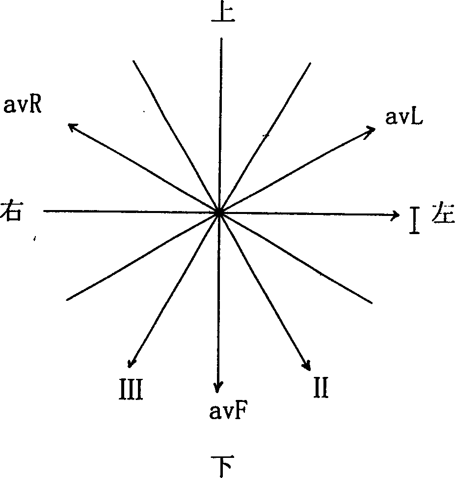
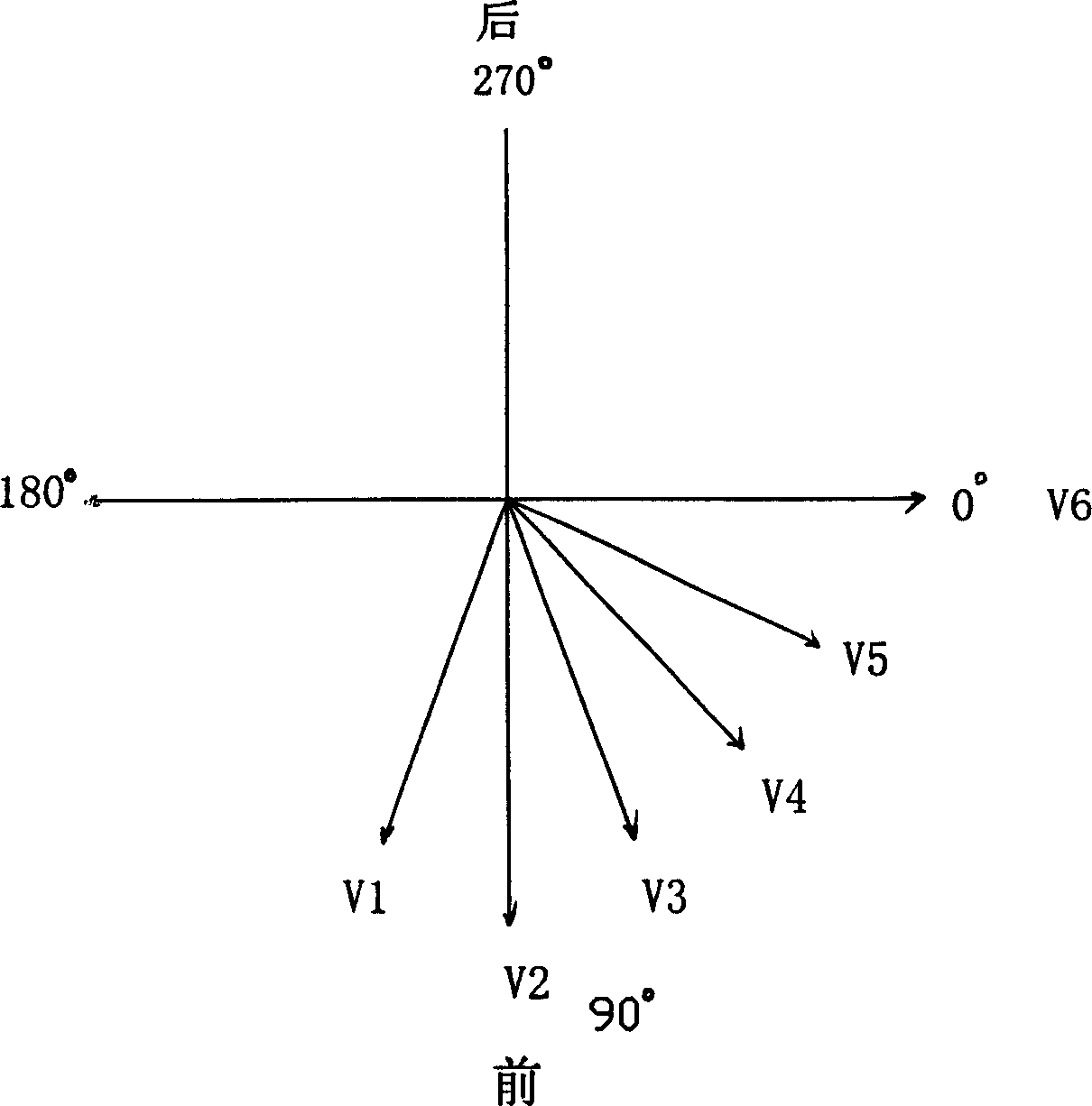
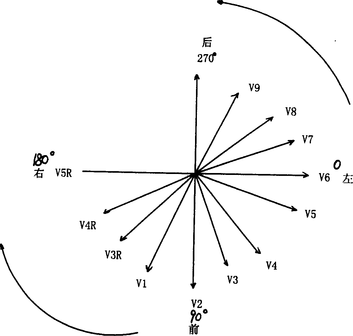
[0088] The present invention converts a variety of synchronous electrocardiogram lead methods in a corrected orthogonal electrocardiogram mode. Orthogonal ECG projection derivation of multiple synchronized ECG lead methods. As shown in Figure 1 and Figure 2, the frontal plane formed by the X-Y axis and the transverse ECG lead axis formed by the X-Z axis are the lead conditions of the current twelve-lead synchronous ECG, which includes three double-leads I, II, and III. Polar limb leads, three unipolar pressurized limb leads avR, avL, and avF, and six chest leads V1-V6. As shown in Figures 3, 4, 5, 6, 7, 8, and 9, on the basis of twelve leads, according to the theory of ECG secondary projection, a dynamic electrocardiogram detector is used. This embodiment uses the BETHUNE-21 series The dynamic electrocardiogram detector realizes multiple synchronous electrocardiogram lead methods of the present invention, and the method is:

[0089] First, the synchronous 18-lead ECG lead me...


PUM


Abstract

The present invention features that the lead method of synchronous 18-lead electrocardiogram converted in corrected orthogonal electrocardiogram mode includes back left deriving V7, V8 and V9 as the three lead axes to reflect the back wall of the left ventricle with V6 lead as base point, right deriving V3R, V4R and V5R as the three lead axes to reflect the right ventricle with V1 lead as base point, and synchronous displaying of the six lead axes and 12 conventional lead axes. The expanded lead method of synchronous 21 leads includes back left deriving V7, V8 and V9 as the three lead axes on the basis of V5 lead axis. The lead method including converting the inclined angles in orthogonal electrocardiogram mode with right and left side correction and multiple bedding plane electrocardiogram lead containing up and down whole angles is the combined application of lateral electrocardiogram and transverse plane/frontal plane electrocardiogram.

Description

technical field [0001] The invention relates to a lead method for electrocardiogram detection instruments used in clinical medicine, especially a hardware system for electrocardiogram amplification in the instrument, which is realized by computer software, and the operation deduced by mathematical derivation method is extremely simple , Only seven body surface electrodes can be used to obtain almost all ECG leads, and a large amount of information can be converted into multiple synchronous ECG lead methods in the form of corrected orthogonal ECG. Background technique [0002] The technical theoretical basis of the present invention originates from the basic theory of ECG vector-orthogonal electrocardiogram and secondary projection of clinical electrocardiogram. [0003] The ECG re-projection theory is the most basic theory of ECG generation, and it is the teaching theory used to explain the generation of ECG in the field of ECG. Its theoretical basis has been recognized by ...

Claims


Application Information

Patent Timeline
Patent Type & Authority Applications(China)
IPC IPC(8): A61B5/0402A61B5/0408
Inventor 段扬段春和
Owner 比顿(北京)医用设备有限公司
Why Eureka
Try Eureka