Inhibition of ferrochelatase as an antiangiogenic therapy

a technology of ferrochelatase and antiangiogenic therapy, which is applied in the direction of lyases, enzymology, biochemistry apparatus and processes, etc., can solve the problems of rapid photoreceptor degeneration, fragile new blood vessel formation, and eventual fibrotic scarring

Active Publication Date: 2020-08-25
INDIANA UNIV RES & TECH CORP
View PDF5 Cites 1 Cited by
  • Summary
  • Abstract
  • Description
  • Claims
  • Application Information

AI Technical Summary

Problems solved by technology

After pathological angiogenesis occurs, newly formed blood vessels are fragile, porous and not fully differentiated.
The formation of such new blood vessels in the eye may lead to hemorrhage, rapid photoreceptor degeneration, and eventual fibrotic scarring, with rapid, permanent vision loss.
AMD has an estimated loss of productivity burden of 55.4 billion annually in the United States.
Severely affected patients have a very poor quality of life, comparable to that of catastrophic stroke victims or advanced cancer patients in constant pain.
Blinding intraocular side effects are also possible and the long-term risks of these drugs are still unclear.
Moreover, because they are biologics, the cost-benefit ratios of these drugs are unfavorable.
For instance, ranibizumab costs approximately $2,000 per monthly dose, rendering these treatments unaffordable for many patients.
VEGF is essential for signaling normal vessel growth during development, but when aberrantly expressed at high levels, causes improper neovessel growth.
Neovessels, extending into the vitreous, do not oxygenate the retina well and easily rupture, leading to retinal ganglion cell and photoreceptor loss, retinal detachment, and blindness.
Although biologic treatments are effective for retinopathy of prematurity and show fewer side effects than surgical treatments, there remain significant concerns about lasting toxic or developmental effects in neonates, especially since these drugs can have systemic actions even when delivered locally.

Method used


Image

  • Inhibition of ferrochelatase as an antiangiogenic therapy
  • Inhibition of ferrochelatase as an antiangiogenic therapy
  • Inhibition of ferrochelatase as an antiangiogenic therapy

Examples


examples 1-4

[0037]Materials & Methods

[0038]EBM-2 and IMDM growth media were purchased from Lonza (Walkersville, Md., USA). HRECs and Attachment Factor were purchased from Cell Systems (Kirkland, Wash., USA). Clonetics® HUVECs were purchased from Lonza. All endothelial cells were used between passages 5 and 8. Endothelial Growth Medium (EGM-2) was prepared by mixing the contents of an EGM-2 “Bullet Kit” (Cat no. CC-4176) with Endothelial Basal Medium (EBM) (Lonza). 92-1 and ARPE-19 cells were grown in RPMI and DMEM media supplemented with 10% FBS and 1% penicillin-streptomycin as described in Basavarajappa et al. J Med Chem 58, pp. 5015-5027 (2015); identity was confirmed by STR profiling. Click-iT TUNEL Alexa Fluor-594 imaging assay kit (Cat no. C10246) was purchased from Molecular Probes (Eugene, Oreg., USA). Monoclonal antibody against α-tubulin (DM1A), protoporphyrin 5-aminolevulinic acid, hemin, griseofulvin and L-arginine were purchased from Sigma-Aldrich (St. Louis, Mo., USA). N-methyl pr...

example 1

[0079]In this Example, protein modulators of angiogenesis were identified.

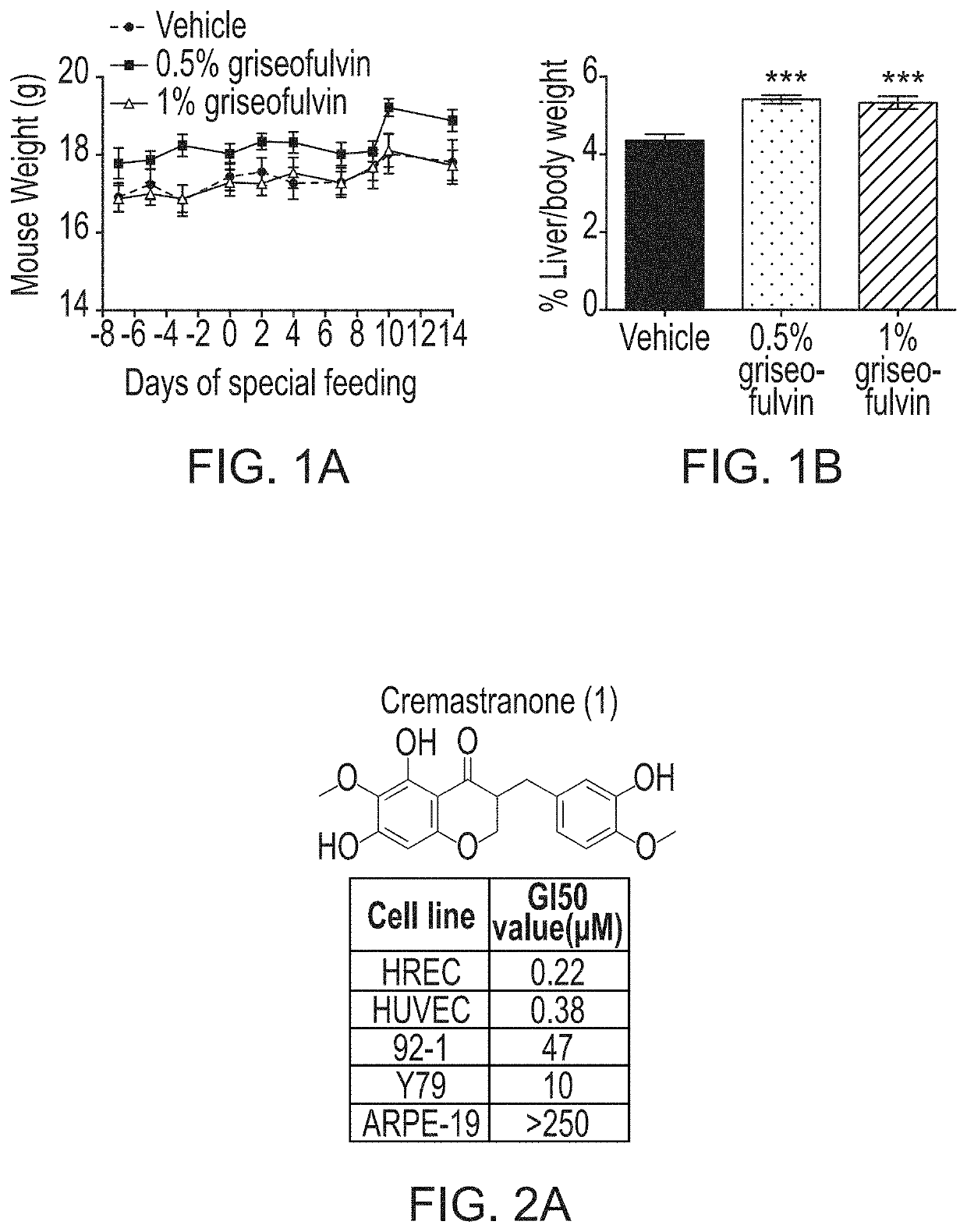
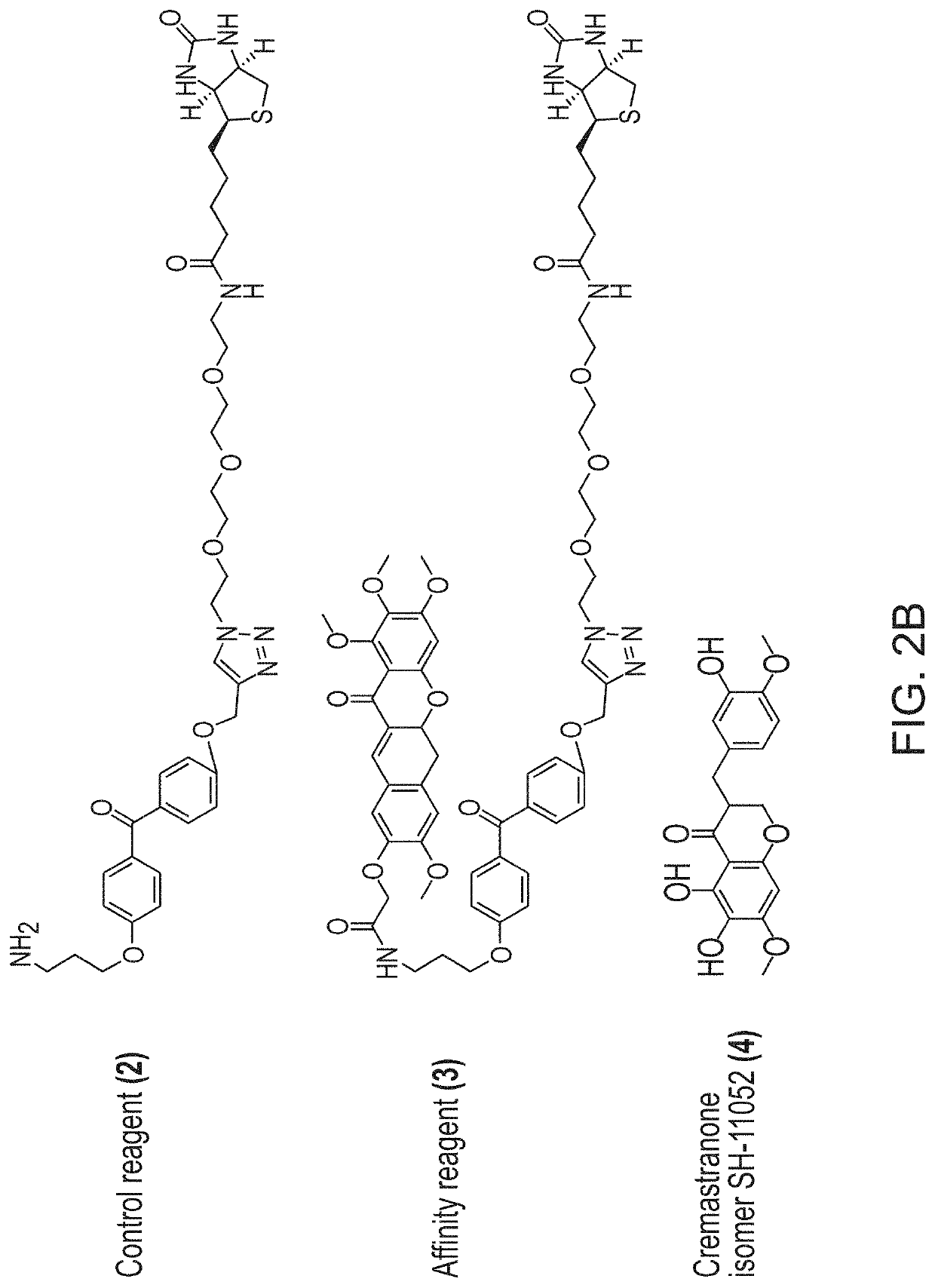
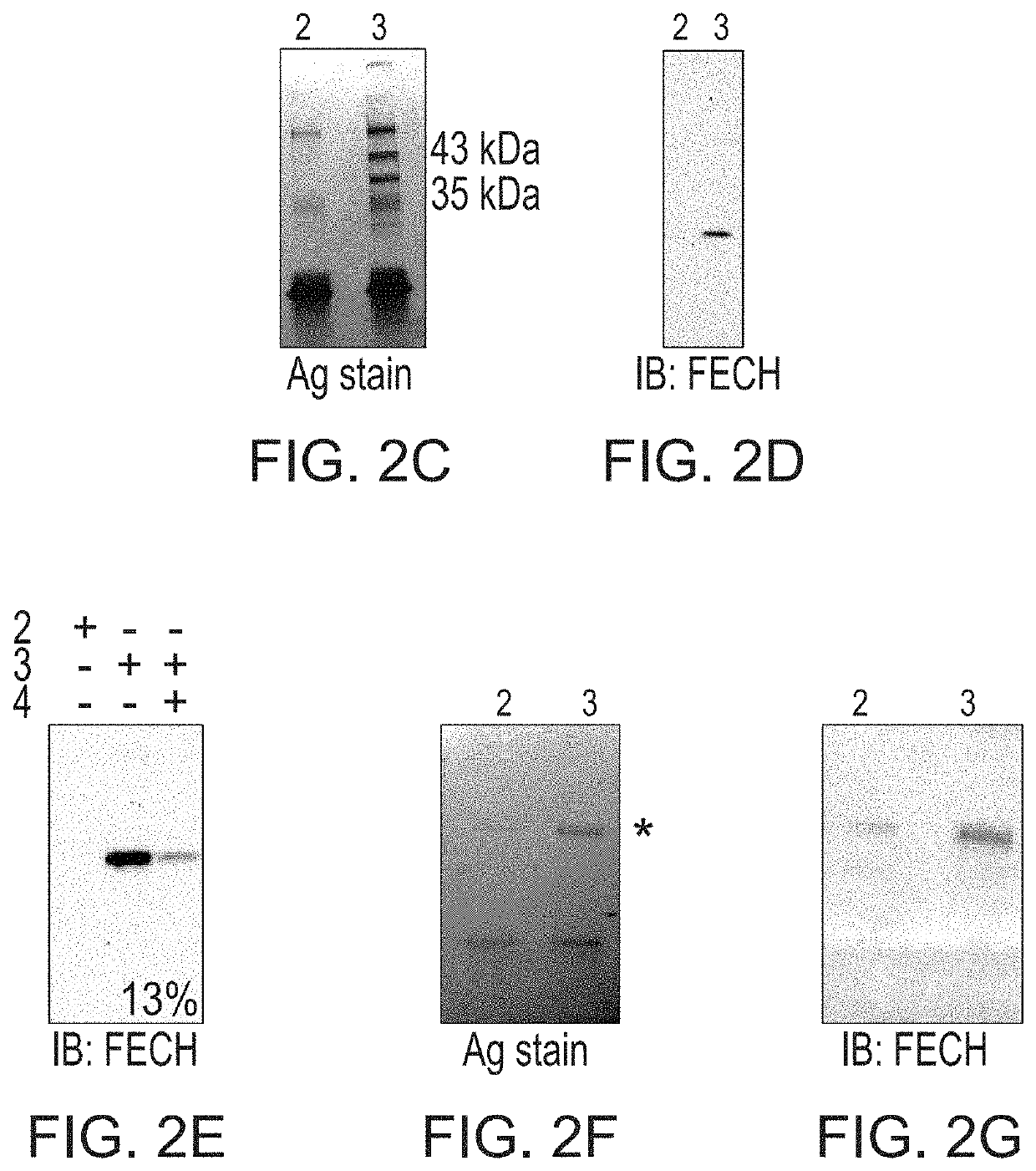
[0080]Photoaffinity chromatography was used to search for targets of the naturally occurring antiangiogenic compound, cremastranone (FIG. 2A), which has selective antiproliferative effects on endothelial cells. Protein binding partners of cremastranone were pulled down from a tissue lysate using immobilized affinity reagent, but not a negative control reagent (FIG. 2B). One of the two pulled down proteins was identified using peptide mass fingerprinting as ferrochelatase (FIG. 2C and Table 1) Immunoblot of eluates from photoaffinity pull down experiments confirmed the identity of the pulled down protein using an antibody against ferrochelatase (FIG. 2D). In order to confirm specificity of binding between cremastranone and pulled down proteins, affinity reagent was incubated with tissue proteins in the presence of excess active cremastranone isomer (FIG. 2B). Under this condition, the binding of ferrochelatase ...

example 2

[0084]In this Example, a known pharmacological inhibitor of ferrochelatase was analyzed for its antiangiogenic properties in vitro.

[0085]N-methyl protoporphyrin (NMPP), a competitive inhibitor of FECH activity, also inhibited proliferation, migration and tube formation ability of HRECs in vitro (FIGS. 5A-5C). However, despite these potent antiproliferative effects, FECH knockdown and low-dose chemical inhibition were not associated with increased apoptosis of these cells (FIG. 6), indicating a cytostatic rather than cytotoxic effect. Moreover, FECH knockdown did not inhibit proliferation of non-endothelial ocular cell lines 92-1 and ARPE-19 as well as macrovascular endothelial cell HUVECs (FIGS. 7A-7C), indicating that FECH inhibition is not associated with ocular cytotoxicity. Together, these experiments confirmed that ferrochelatase function is required for angiogenesis in vitro.


PUM

PropertyMeasurementUnit
pHaaaaaaaaaa
pHaaaaaaaaaa
pHaaaaaaaaaa

Abstract

Methods for treating angiogenesis-mediated diseases are disclosed. More particularly, the present disclosure relates to methods of inhibiting ferrochelatase as an antiangiogenic therapy.

Description

CROSS REFERENCE TO RELATED APPLICATIONS[0001]This application claims priority to U.S. Provisional Application No. 62 / 111,149 filed on Feb. 3, 2015, which is hereby incorporated by reference in its entirety.BACKGROUND OF THE DISCLOSURE[0002]The field of the disclosure relates generally relates to methods for treating angiogenesis-mediated diseases. More particularly, the present disclosure relates to methods of inhibiting ferrochelatase as an antiangiogenic therapy.[0003]Angiogenesis does not occur in the body, except during development and wound repair processes. However, during numerous pathological conditions, angiogenesis occurs, notably in ocular diseases such as retinopathy of prematurity (ROP), diabetic retinopathy (DR), and “wet” age-related macular degeneration (AMD). After pathological angiogenesis occurs, newly formed blood vessels are fragile, porous and not fully differentiated. The formation of such new blood vessels in the eye may lead to hemorrhage, rapid photorecepto...

Claims


Application Information

Patent Timeline
Patent Type & Authority Patents(United States)
IPC IPC(8): C12N15/11A61K45/06A61K9/00A61K31/713A61K31/343C12N15/113A61K31/409
CPCA61K45/06C12N15/1137C12Y499/01001A61K9/0019A61K31/409A61K31/343A61K31/713C12N2310/14
Inventor CORSON, TIMOTHYBASAVARAJAPPA, HALESHA
Owner INDIANA UNIV RES & TECH CORP
Why Eureka
Try Eureka