Method to detect molecular binding by surface-enhanced Raman spectroscopy

Inactive Publication Date: 2005-10-06
INTEL CORP
View PDF10 Cites 20 Cited by
  • Summary
  • Abstract
  • Description
  • Claims
  • Application Information

AI Technical Summary

Problems solved by technology

However, for many analytes the requirements for analytical and functional sensitivity are becoming more and more demanding.
However, conventional methods are often affected by strong background signal due to non-specific binding, which generates misleading information about the binding eve

Method used


Image

  • Method to detect molecular binding by surface-enhanced Raman spectroscopy
  • Method to detect molecular binding by surface-enhanced Raman spectroscopy
  • Method to detect molecular binding by surface-enhanced Raman spectroscopy

Examples


example 1

SERS Detection of Unlabeled Antibody

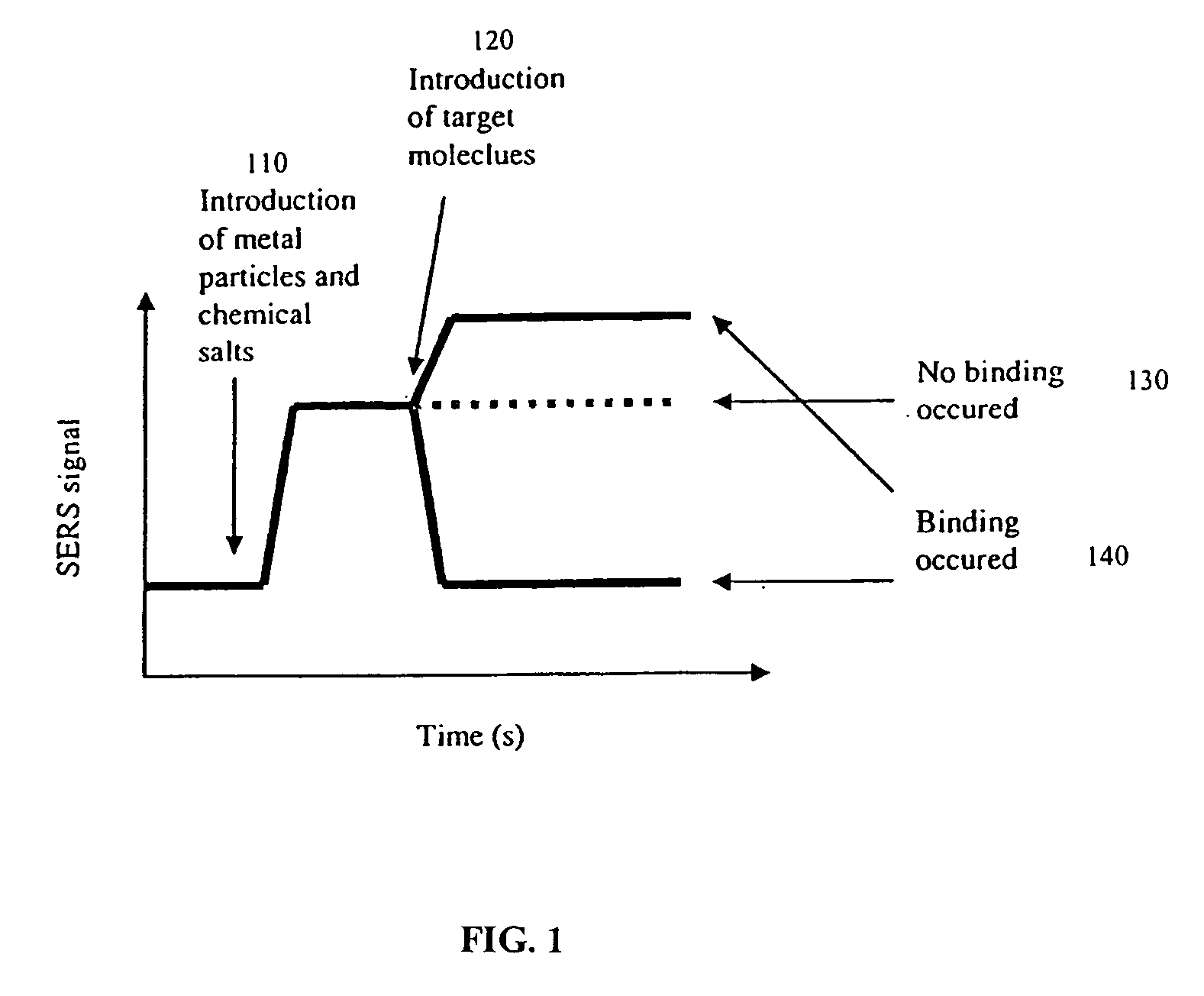
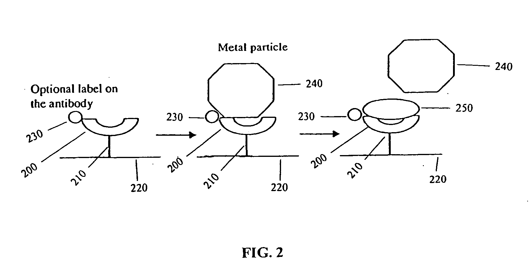
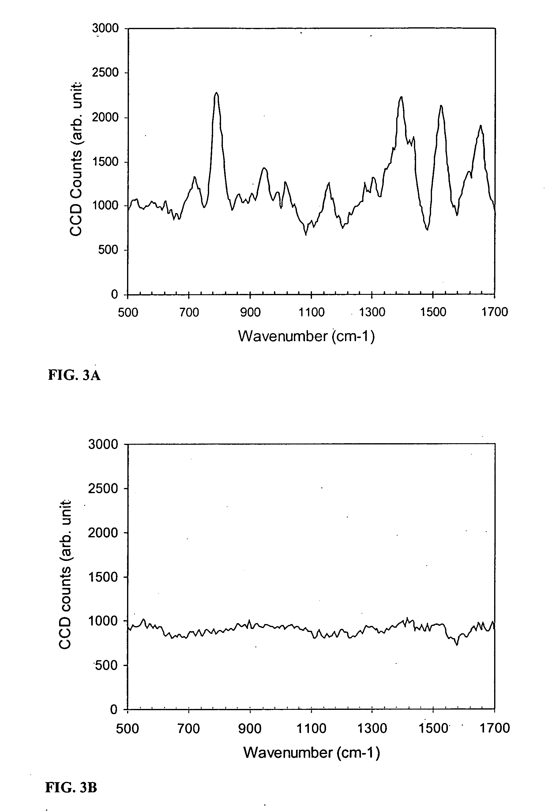
[0104] This example illustrates the detection of an antibody using SERS. Antibody molecules were immobilized on the gold-coated substrate by using EDC chemistry (1-Ethyl-3-[3-dimethylaminopropyl]carbodiimide Hydrochloride), developed by and available from Pierce (Rockford, Ill.). The control was a blank substrate with EDC treatment and no antibody. Eighty microliters of a mixture of colloidal silver (synthesized by the recipe published by Lee and Meisel, J. Phys. Chem. 1982, 986, 3391-3396) and lithium chloride salt solution were applied onto the sample and the control before spectrum collection.

[0105] A Raman microscope was used to collect the spectrum from the antibody sample and the control sample. The Raman microscope included an argon ion laser (Coherent, Santa Clara, Calif.), an optical microscope (Nikon), optical filters (Kaiser Optical, Ann Arbor, Mich.), a spectrograph (Acton Research, Acton, Mass.), and a CCD camera (Roper Scientific, ...


PUM


Abstract

Provided herein are methods for detecting molecular binding using surface enhanced Raman spectroscopy (SERS). The SERS signal can be generated by associating one of the binding partners with a SERS-active particle or substrate. Binding is detected by detecting a change in a SERS signal after two binding partners are contacted with each other as compared to before the binding partners are contacted with each other. The method is useful for detecting binding of biomolecules such as antibodies to antigens and receptors to ligands.

Description

BACKGROUND OF THE INVENTION [0001] 1. Field of the Invention [0002] The invention relates generally to detection and analysis of biomolecules, and more specifically to detection of binding between biomolecules. [0003] 2. Background Information [0004] Molecular binding assays, such as immunoassays, are frequently used for the detection of analytes in serum, plasma, urine or other body fluid samples for medical and diagnostic purposes. A plethora of analytes are detectable by molecular binding assays. However, for many analytes the requirements for analytical and functional sensitivity are becoming more and more demanding. [0005] Conventionally, molecular binding is observed by detecting fluorescent or radioactive labels on either an antibody or a target molecule. However, conventional methods are often affected by strong background signal due to non-specific binding, which generates misleading information about the binding events or limits the functional sensitivity of the method. Fo...

Claims


Application Information

Patent Timeline
IPC IPC(8): G01N21/65G01N33/543G01N33/553
CPCG01N21/658G01N33/54373G01N33/553
Inventor KOO, TAE-WOONGCHAN, SELENA
Owner INTEL CORP
Why Eureka
Try Eureka