System and method for providing communication between ultrasound scanners

a technology of ultrasound scanner and communication system, applied in the field of medical imaging system, can solve the problems of requiring more processing power and large amounts of raw data to process this more complex raw data

Active Publication Date: 2006-01-12
GENERAL ELECTRIC CO
View PDF17 Cites 28 Cited by
  • Summary
  • Abstract
  • Description
  • Claims
  • Application Information

AI Technical Summary

Problems solved by technology

Moreover, output displayed as three dimensional (3D) and time varying 3D images (known as 4D images) also require larger amounts of raw data and additional processing power to process this more complex raw data.
However, this will require modifying existing scanner, replacing existing scanners or performing scans in steps if the processing power of the scanner is insufficient to process the data in a single process.

Method used


Image

  • System and method for providing communication between ultrasound scanners
  • System and method for providing communication between ultrasound scanners
  • System and method for providing communication between ultrasound scanners

Examples


Embodiment Construction

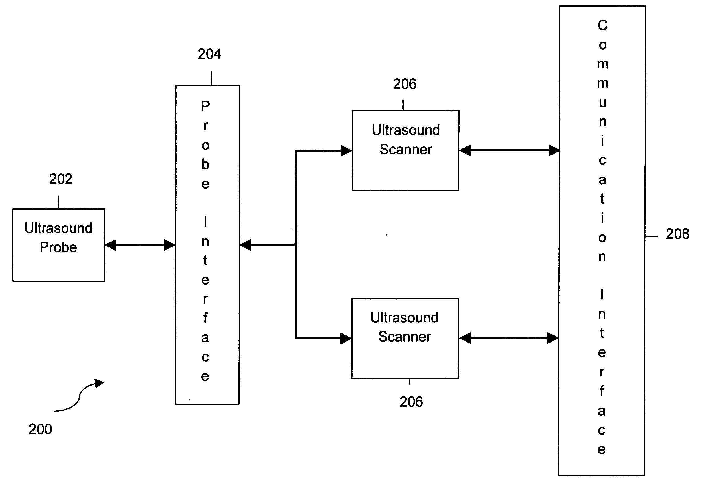
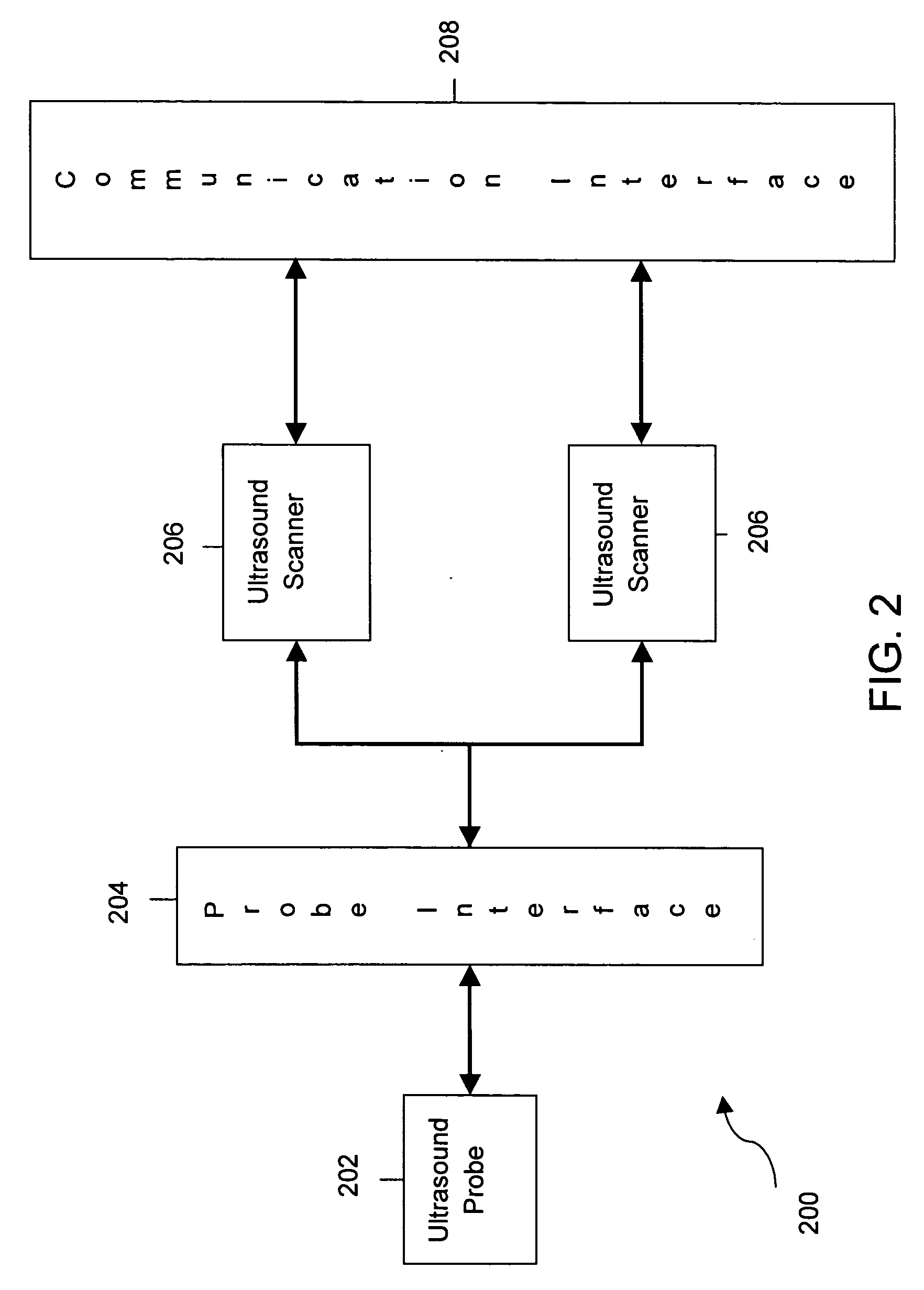
[0013] Various embodiments of the invention provide a system and method for combining medical imaging devices. The embodiments utilize parallelism in an application flow where the application requires high computational resources. Multiple channels of data flow into the system via a transducer element, with different portions of the multiple channels of data processed by different scanners.

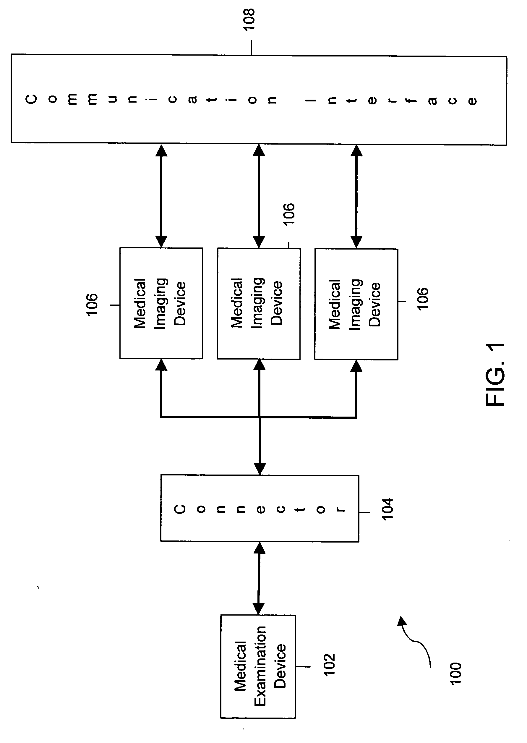
[0014] Specifically, FIG. 1 is a block diagram of a medical imaging system in accordance with an exemplary embodiment of the invention. Medical imaging system 100 includes a medical examination device 102 to collect raw input data. The data can be data such as, for example, echoes from an ultrasound probe or data for an electrocardiograph (ECG). Medical examination device 102 may be any medical examination device such as, for example, an ultrasound probe or an electroencephalogram (EEG) needle. Medical examination device 102 is connected via a connector 104 to a plurality of medical imaging devic...


PUM


Abstract

System and method for ganged ultrasound scanners is described. The described system includes a plurality of ultrasound scanners configured as a network and at least one connector for connecting a single ultrasound probe to the plurality of ultrasound scanners. The connector is configured to provide communication between the plurality of ultrasound scanners and the ultrasound probe.

Description

BACKGROUND OF THE INVENTION [0001] This invention relates generally to medical imaging systems, and more particularly, to providing communication between ultrasound scanners in a medical imaging system. [0002] Medical imaging systems are requiring more computational power and memory to perform scans providing higher quality images. Particularly in ultrasound diagnostic medical imaging, the imaging processes are requiring higher computational processing power. The processes require more CPU, memory and storage capacity to perform the image processing required in ultrasound imaging. Improved image quality require larger data sizes to be processed. Moreover, output displayed as three dimensional (3D) and time varying 3D images (known as 4D images) also require larger amounts of raw data and additional processing power to process this more complex raw data. [0003] To provide improved quality images, probes or other medical examination devices must be configured to acquire larger sizes o...

Claims


Application Information

Patent Timeline
Patent Type & Authority Applications(United States)
IPC IPC(8): A61B8/00
CPCA61B8/00A61B8/4411G06F19/3418A61B8/56A61B8/463A61B8/44G16H40/67G16H30/20
Inventor MATHEW, PRAKASH PARAYILCHILUKURI, SASTRY V.S.
Owner GENERAL ELECTRIC CO
Why Eureka
Try Eureka