

Angiographic x-ray diagnostic device for rotation angiography
- Summary
- Abstract
- Description
- Claims
- Application Information
AI Technical Summary
Benefits of technology
Problems solved by technology
Method used
Image

Examples
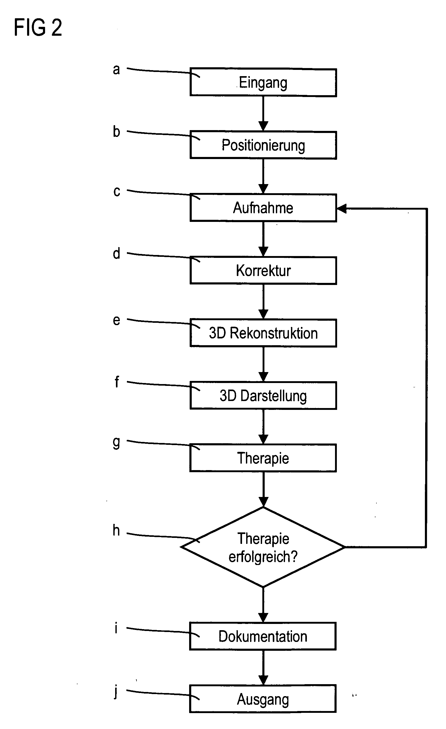
Embodiment Construction
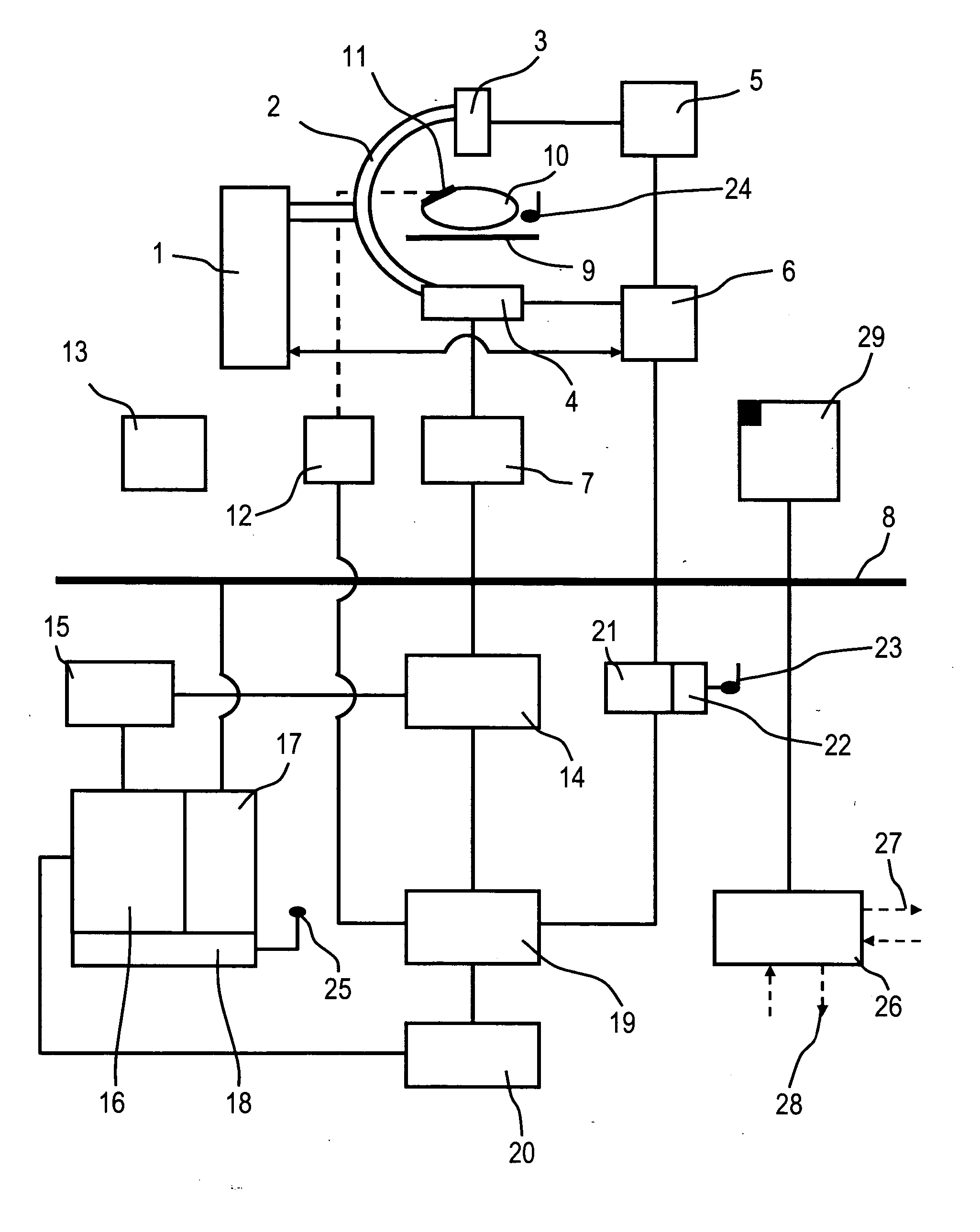
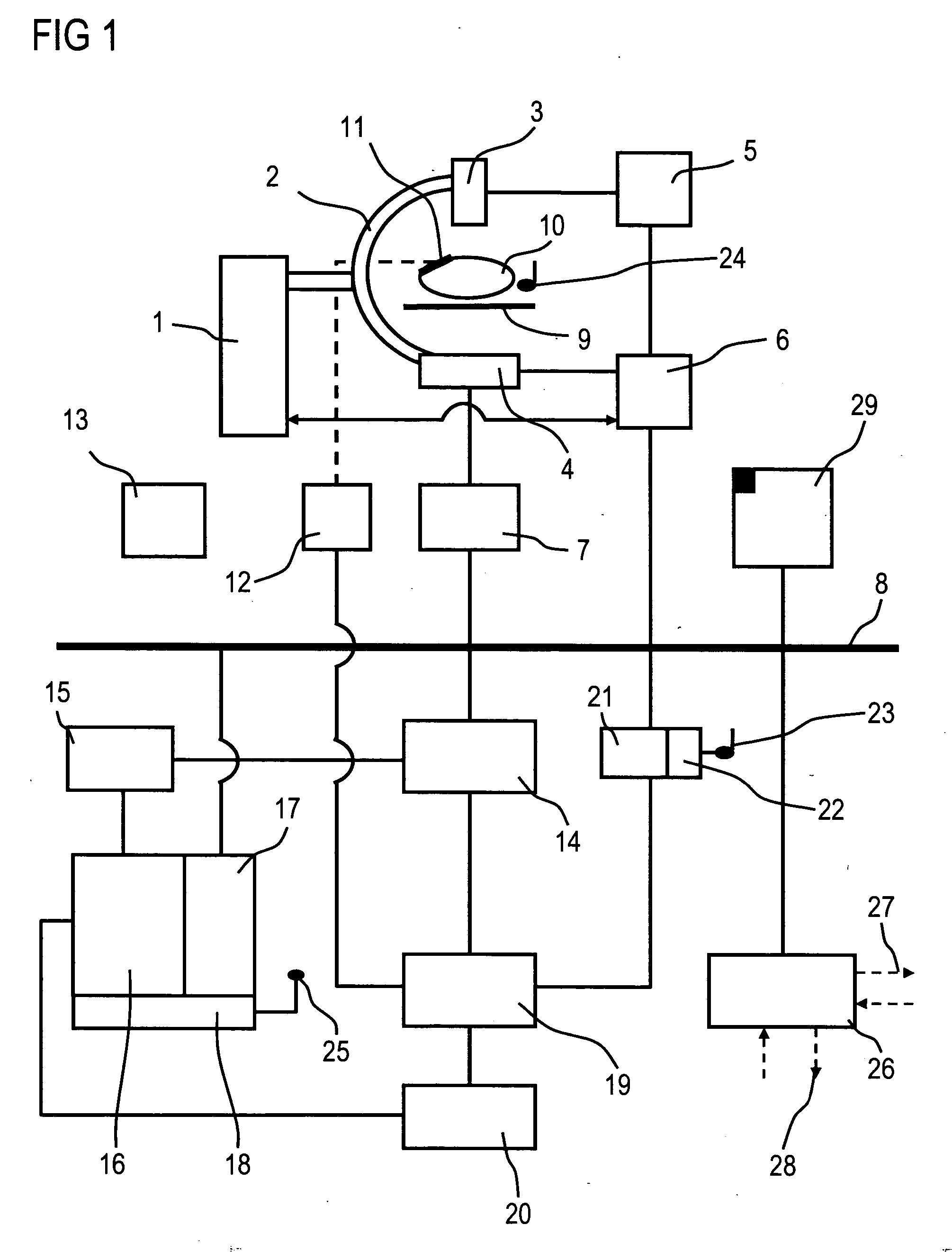
[0020]FIG. 1 shows an x-ray diagnostic device comprising a C-arm 2 which is mounted in a rotatable manner on a stand 1, at the end of which is mounted an x-ray emitter 3 and an x-ray image detector 4.
[0021] Instead of the support 1 displayed, floor and / or ceiling supports can also be used. The C-arm 2 can also be replaced by a so-called electronic C-arm 2, thereby effecting an electronic coupling of the x-ray emitter 3 and x-ray image detector 4, which causes a circular path to be traveled from the x-ray emitter 3 and the x-ray image detector 4, controlled by a computing unit for instance.
[0022] The x-ray image detector 4 can be a flat, rectangular and / or square semiconductor detector which is preferably created from amorphous silicon (aSi).
[0023] A high voltage generator 5 is connected to a system controller 6 and drives the x-ray emitter 3. The system controller 6 is furthermore connected to the x-ray image detector 4, for instance the aSi flat detector, for the synchronous con...
PUM
 Abstract
Description
Claims
Application Information