Diversion Of Emboli During Fluid Circulation

a technology of fluid circulation and embolism, which is applied in the direction of suction devices, valves, haemofiltration, etc., can solve the problems of increased risk of gaseous emboli, increased risk of embolism, and unobtrusive formation of blood clots and gas bubbles

Inactive Publication Date: 2008-08-28
SIFR2000
View PDF13 Cites 2 Cited by
  • Summary
  • Abstract
  • Description
  • Claims
  • Application Information

AI Technical Summary

Problems solved by technology

During extracorporeal circulation in e.g. heart surgery there is a risk to introduce detrimental matter into the circulation of the patient.
The formation of blood clots and gas bubbles can occur unobtrusively despite rigorous routines to avoid the formation of these emboli, or it can occur from malpractice by the involved personnel.
In the case the extracorporeal circuit contains a gas-exchange device e.g. an oxygenator there is even a greater risk of gaseous emboli.
It is obvious for a person skilled in the art that bubbles or other emboli of millimeter-size may be harmful for the organism since capillaries are much smaller with diameters a hundred times less.
Thus, the built-in security measures against bubble injection in heart-lung machines currently on the market seem inadequate.
Besides, when an embolus halts the heart-lung machine the patient is deprived completely of circulation and a series of maneuvers has to be performed by the perfusionist and the surgeon to rid the tubing from the embolus.
This may take some time and there is a great risk in misunderstandings and mishandling of the situation due to stress.
Thus, not only the adverse effects of bubbles per se, but also the cumbersome mandatory maneuvers to rid the device of sensed bubbles before their entry into the patient, may be deleterious.
Furthermore, symptoms also include personality changes, difficulties in concentration and emotional unstableness.
Bearing in mind that more than a million extracorporeal procedures are performed in the world annually this is a formidable problem.
The capillary bed of the brain serves as a filter for the blood from the heart-lung machine, which is disadvantageous and at least in part may explain the frequent clinical neurological deficiencies encountered after heart surgery.
None of these methods have means to detect bubbles in the blood stream when they occur.
Also, in catastrophic events, when large amounts of air enter the system, these methods easily get saturated, letting air through into the blood vessels of the patient.

Method used


Image

  • Diversion Of Emboli During Fluid Circulation
  • Diversion Of Emboli During Fluid Circulation
  • Diversion Of Emboli During Fluid Circulation

Examples


first embodiment

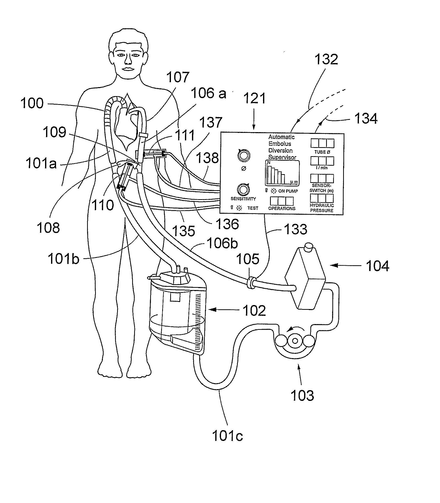
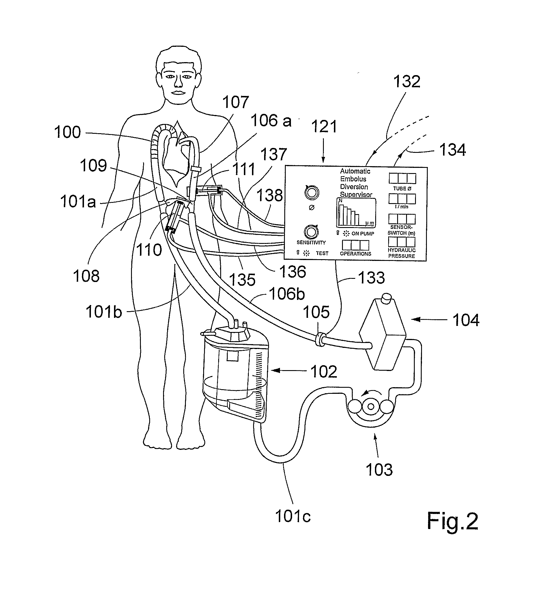
[0025]FIG. 2 shows schematically the present invention. A first fluid circuit 100, 101a, 101b, 101c, 102, 103, 104, 106a, 106b, 107, 109 is established between an outlet from a vascular system of a living being and an inlet into said vascular system. As exemplified in FIG. 2, the outlet is realized as an outlet from the vena cava and the heart and the inlet is realized as an inlet into ascending aorta of the patient. Blood can be extracted from the outlet by means of a extraction means, e.g. a cannula, 100 and extracorporeally circulated in said fluid circuit between the outlet and the inlet.

[0026]A second fluid circuit 101b, 101c, 102, 103, 104, 106b, 108, 109 is configured to bypass the inlet and the outlet of the vascular system. As shown in FIG. 2, the body will thus be bypassed by the second fluid circuit. However, it should be understood that the inlet and outlet do not have to be an inlet and an outlet of the same organ or body part, but they can be an inlet and an outlet of ...

third embodiment

[0040]the inventive system is depicted in FIG. 8. In this application the invention is modified to be used in dialysis. The oxygenator is in this situation not needed and a dialysis membrane 140 is added. In dialysis, the venous reservoir may be of a smaller size as compared to the size of the venous reservoir in the previously described embodiments. However, in case a large bubble has been detected and is redirected according to the method of the invention, the venous reservoir should contain enough volume to minimize operational arrests. In FIG. 8 the dialysis catheter 141 entering the body is depicted as a two-lumen veno-venous dialysis catheter, but other arrangements of cannulation the vascular system for dialysis are feasible.


PUM


Abstract

A method of diversion of an embolus during fluid circulation in an extracorporeal circuit which includes the steps of establishing a first circuit (100, 101a, 101b, 101c, 102, 103, 104, 106b, 106a, 107) between an outlet of a vascular system and an inlet of said vascular system; establishing a 5 second circuit (101b, 101c, 102, 103, 104, 106b, 108) configured to bypass said inlet and outlet; providing a fluid reservoir (102) a pumping means (103), and a bubble detector (105) in said circuits; detecting by means of said bubble detector (105) an embolus in said first circuit and sending a signal to an automatic embolus diversion supervisor (121) connected to said bubble detector (105); and controlling fluid flow direction means (110, 111) in dependence of the signal from said bubble detector (105); whereby said fluid flow direction means (110, 111) are controlled to redirect a detected embolus to said second circuit.

Description

TECHNICAL FIELD[0001]The present invention relates to diversion of an embolus during fluid circulation. More specifically the present invention relates to the separation of an embolus comprised in a flowing fluid before the flowing fluid reaches a target.BACKGROUND OF THE INVENTION[0002]During extracorporeal circulation in e.g. heart surgery there is a risk to introduce detrimental matter into the circulation of the patient. The formation of blood clots and gas bubbles can occur unobtrusively despite rigorous routines to avoid the formation of these emboli, or it can occur from malpractice by the involved personnel. Gas emboli are formed in circulating blood due to cavitation, temperature gradients, and the surplus amount of gases dissolved in blood. In the case the extracorporeal circuit contains a gas-exchange device e.g. an oxygenator there is even a greater risk of gaseous emboli.[0003]Gas emboli can be detected by different techniques, and all modern heart-lung machines are equ...

Claims


Application Information

Patent Timeline
Patent Type & Authority Applications(United States)
IPC IPC(8): A61M1/34
CPCA61M1/3627A61M1/3626A61M39/223A61M2039/226A61M39/281A61M39/227
Inventor BORIS-MOLLER, FREDRIK
Owner SIFR2000
Why Eureka
Try Eureka