Display of a medical image

a medical image and display technology, applied in the field of display of medical images, can solve the problem that the computer keyboard or the computer mouse cannot be positioned in the immediate service area of the patient positioning device, and achieve the effect of improving the service life and improving the service life of the patien

Inactive Publication Date: 2009-08-27
SIEMENS AG
View PDF7 Cites 92 Cited by
  • Summary
  • Abstract
  • Description
  • Claims
  • Application Information

AI Technical Summary

Benefits of technology

[0006]The present embodiments may obviate one or more of the drawbacks or limitations inherent in the related art. For example, in one embodiment, a device allows displayed image information to be modified in a simple user-friendly manner.

Problems solved by technology

As a result, the computer keyboard or the computer mouse may not be positioned in the immediate service area of a patient positioning device, such as a patient examination table, on which the patient is positioned for the medical examination or intervention.

Method used


Image

  • Display of a medical image
  • Display of a medical image
  • Display of a medical image

Examples


Embodiment Construction

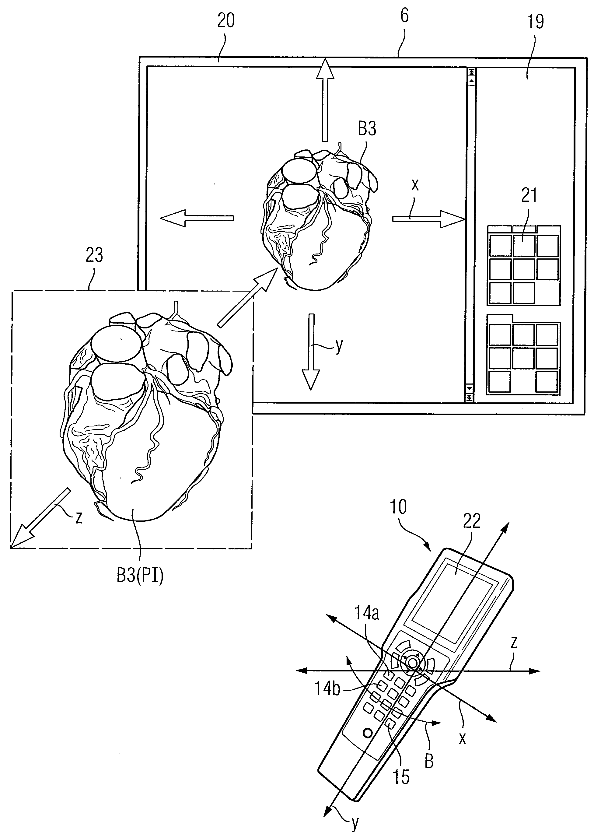
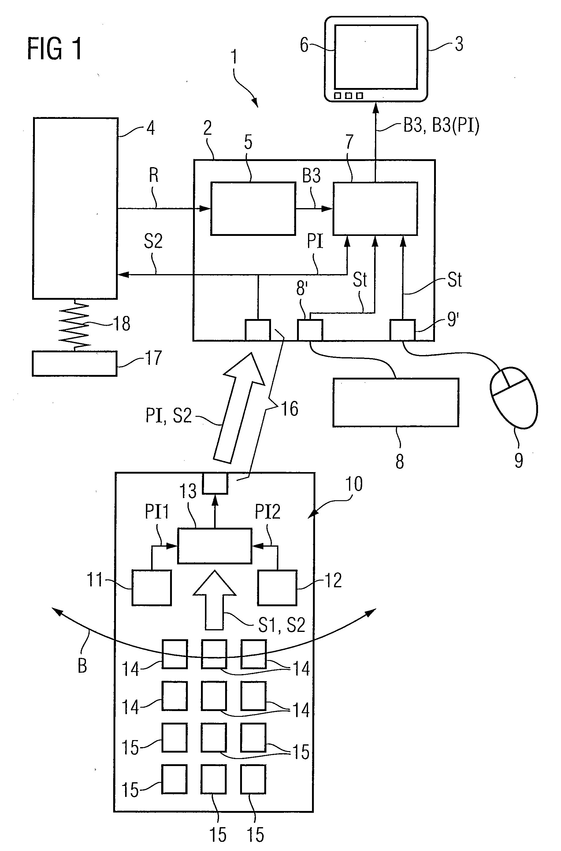
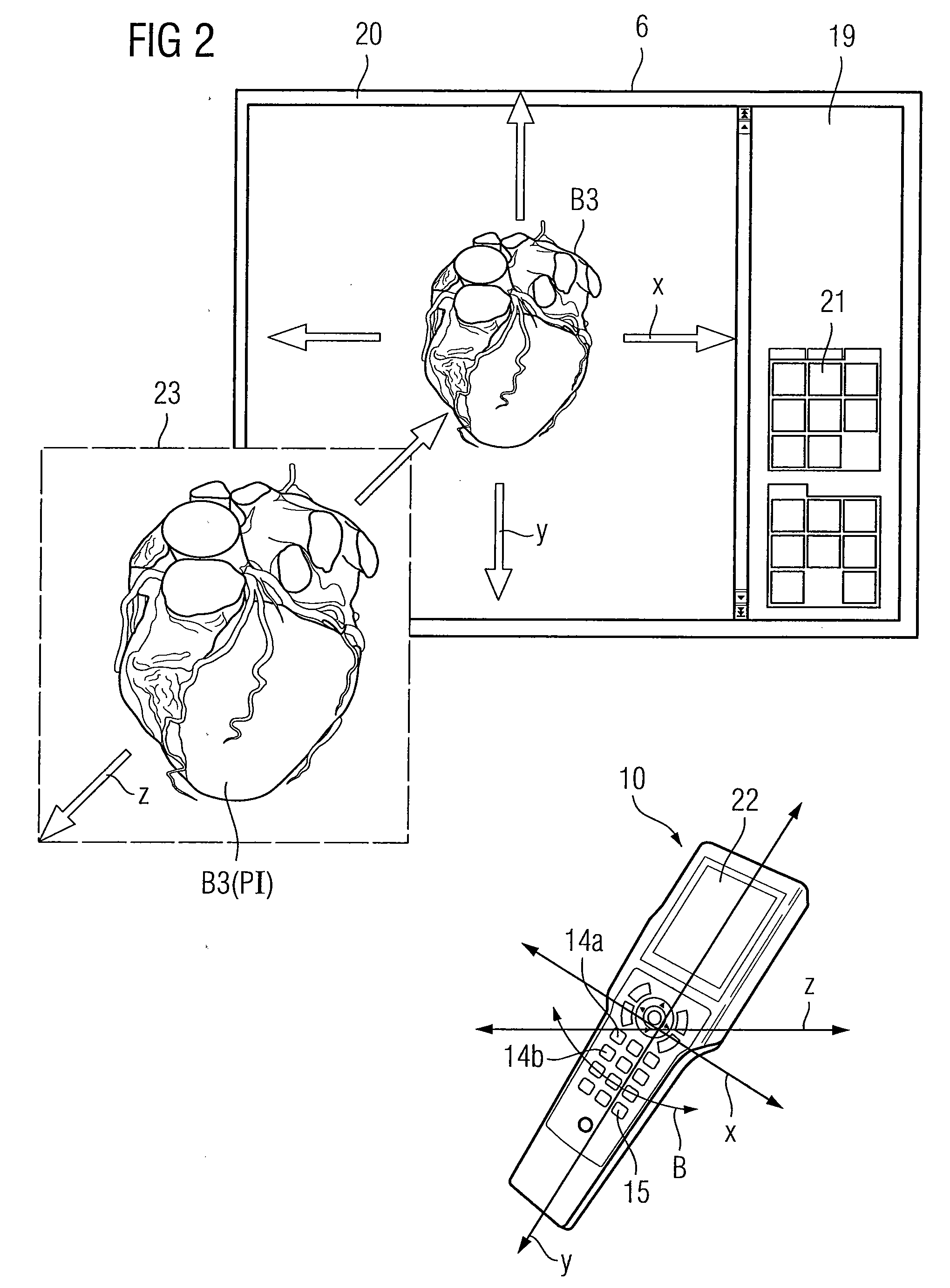
[0035]FIG. 1 shows a device for displaying a three-dimensional medical image 1 with a processing unit 2 taking the form of a computer system and with a display element 3 connected to the processing unit and taking the form of a monitor. The device 1 is associated with a medical imaging system 4 which takes the form, for example, of a computer tomograph (CT), a positron emission tomograph (PET), a single photon emission computed tomograph (SPECT) or a magnetic resonance tomograph (MRT). The medical imaging system 4 raw data R is measured. The raw data R is processed by an arithmetic unit 5 associated with the processing unit 2. A three-dimensional medical image B3 is computed and displayed on a display 6 of the display element 3. In the case of a computer tomograph, projection images are measured as raw data R, which is converted by an arithmetic unit 5, taking the form of a reconstruction computer into a three-dimensional medical image B3. A software module 7 is provided for the pre...


PUM


Abstract

A device for displaying a medical image is provided. The device includes a processing unit, a display, a remote control, a communication interface, and a software module. The processing unit is operable to process the medical image information. The display is operable to display the medical image information. The remote control is operable to register a user movement by at least one motion-sensitive sensor. The communication interface is operable to transfer the user movement to the processing unit. The software module is associated with the processing unit. The software module is operable to reconcile the user movement with the medical image information so that the user movement is reproduced as a virtual movement of the displayed medical image information.

Description

[0001]This patent document claims the benefit of DE 10 2008 010 717.4, filed Feb. 22, 2008, which is hereby incorporated by reference.BACKGROUND[0002]The present embodiments relate to displaying a medical image. In particular, the present embodiments relate to a device, method, and imaging system for displaying a medical image.[0003]Imaging systems are available for generating medical image information. The imaging systems include for example a Computer Tomograph (CT), a Positron Emission Tomograph (PET), a Single Photon Emission Computed Tomograph (SPECT) or a Magnetic Resonance Tomograph (MRT). The imaging system is used to obtain an image of a body region of a person. Medical image information includes image information that can be obtained using the imaging system. The image in question is normally a two-dimensional (2D), three-dimensional (3D) or four-dimensional (4D) image. A two-dimensional image encompasses two spatial dimensions. A three-dimensional image encompasses three ...

Claims


Application Information

Patent Timeline
Patent Type & Authority Applications(United States)
IPC IPC(8): G06F3/048G01P15/00G06F3/033G06F3/0346G06F3/0484
CPCG06F3/0346A61B6/548G06F3/04845
Inventor KAGERMEIER, ROBERTFIETZEL, EIKESCHROTER, STEFFENSIERK, DIETMAR
Owner SIEMENS AG
Why Eureka
Try Eureka