Systems and Methods for Automatic Vertebra Edge Detection, Segmentation and Identification in 3D Imaging

a technology of automatic vertebrae edge detection and 3d imaging, applied in image analysis, image enhancement, instruments, etc., can solve the problems of insufficient accuracy, inaccurate segmentation and identifying of vertebrae, and feature-based recognition techniques can suffer from accuracy problems, and achieve the effect of high fit respons

Active Publication Date: 2011-03-10
SIEMENS HEALTHCARE GMBH
View PDF7 Cites 48 Cited by
  • Summary
  • Abstract
  • Description
  • Claims
  • Application Information

AI Technical Summary

Benefits of technology

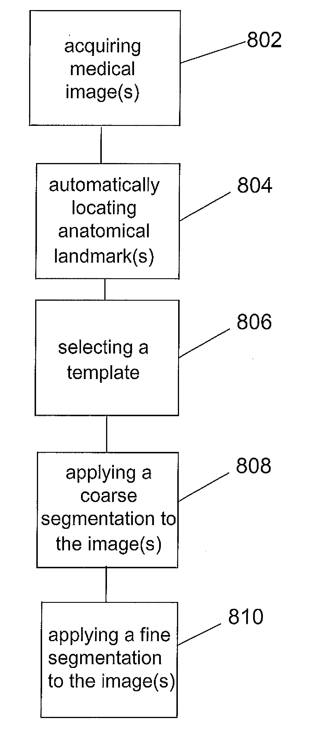
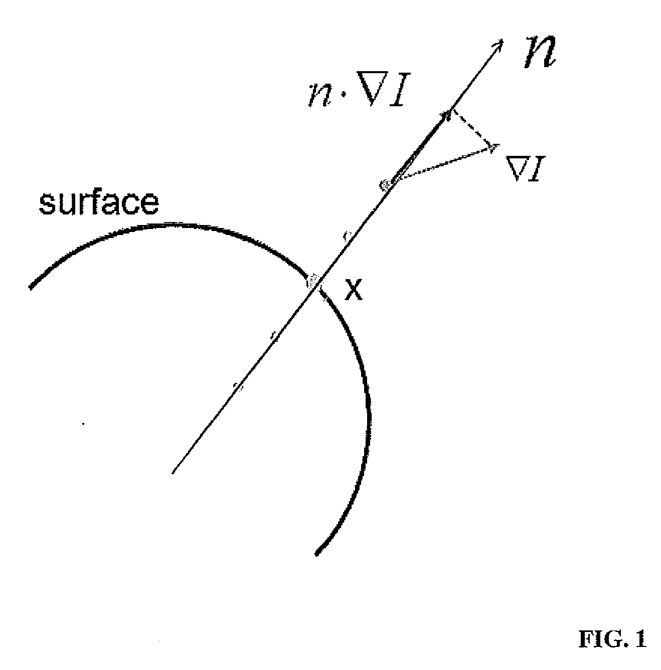
[0012]According to an aspect of the present disclosure, a system for automated segmentation of anatomical structures in image data is provided, including a digital image input for receiving at least one digital image of an anatomical structure, a storage device, coupled to the image input for storing the at least one digital image, and a processor coupled to the storage device. The processor being configured to receive the at least one digital image, analyze the at least one digital image and automatically locate at least one anatomical landmark, select a template from a database of pre-defined templates based upon the at least one anatomical landmark, and segment the at least one digital image by applying a coarse segmentation and a fine segmentation to obtain an optimized segmented template. Applying the coarse segmentation comprises, for each of a plurality of sub-regions of the anatomical structure, applying a corresponding sub-region of the selected template to the anatomical structure, calculating a first edge response, deforming the sub-region of the selected template, calculating a deformation edge response, selecting the sub-region or deformed sub-region of the template having a highest edge response, and combining the highest edge-response sub-region from each of the plurality of sub-regions into a single coarse-segmented template. Applying the fine segmentation comprises, for each of a plurality of patches of the coarse-segmented template, calculating an edge-response of the patch relative to the anatomical structure, deforming the patch, calculating an edge-response of the deformed patch, selecting the patch or deformed patch having a highest edge-response, and combining the highest edge-response patches into a single optimized segmented template. The digital image can be at a three-dimensional (3D) image, and the template can be a 3D surface template. Receiving the digital image can include acquiring the image using an imager selected from: computer tomography (CT), positron emission tomography (PET), single photon emission computed tomography (SPECT), magnetic resonance (MR), and ultrasound. Selecting a template can include selecting a template from a database of pre-defined templates based upon the at least one anatomical landmark, performing a plurality of rigid transformation on the selected template, calculating a fit response for each transformed template, and selecting a transformed template having a highest fit response. Deforming the template can include performing a plurality of translations of the template along each of the x, y, and z axes, performing a plurality of re-scalings of the template along each of the x, y, and z axes, and performing a plurality of rotations of the template along each of the x, y, and z axes. Subsequent to combining the highest edge-response sub-region from each of the plurality of sub-regions, the processor can perform a smoothing operation to smooth the single coarse-segmented template. Deforming the patch can include calculating a mean normal for the patch, and deforming the patch along the mean normal. Subsequent to combining the highest edge-response patches, the processor can perform a smoothing operation to smooth the single optimized segmented template.

Problems solved by technology

Recognizing anatomical structures within digitized medical images presents multiple challenges.
However, feature based recognition techniques can suffer from accuracy problems.
Unfortunately, the task of segmenting and identifying vertebrae, even using CAD systems that are well-developed for other anatomical structures, often proves inaccurate and therefore inadequate.
The difficulty lies in the inherent complexity of vertebrae.
However, the variation within the same class of vertebra as well as the variation in neighboring structures makes vertebral modeling and imaging extremely difficult.
However, that identification algorithm is based on vertebral appearance model (i.e., average volume block) spatial registration and matching which is extremely computationally consuming.
In a real-world hospital setting, such system dedication to a single patient's data is not practical or realistic.

Method used


Image

  • Systems and Methods for Automatic Vertebra Edge Detection, Segmentation and Identification in 3D Imaging
  • Systems and Methods for Automatic Vertebra Edge Detection, Segmentation and Identification in 3D Imaging
  • Systems and Methods for Automatic Vertebra Edge Detection, Segmentation and Identification in 3D Imaging

Examples


Embodiment Construction

[0027]In the following description, numerous specific details are set forth such as examples of specific components, devices, methods, etc., in order to provide a thorough understanding of embodiments of the present invention. It will be apparent, however, to one skilled in the art that these specific details need not be employed to practice embodiments of the present invention. In other instances, well-known materials or methods have not been described in detail in order to avoid unnecessarily obscuring embodiments of the present invention. While the invention is susceptible to various modifications and alternative forms, specific embodiments thereof are shown by way of example in the drawings and will herein be described in detail. It should be understood, however, that there is no intent to limit the invention to the particular forms disclosed, but on the contrary, the invention is to cover all modifications, equivalents, and alternatives falling within the spirit and scope of th...


PUM


Abstract

Systems and methods for automatic accurate and efficient segmentation and identification of one or more vertebra in digital medical images using a coarse-to-fine segmentation.

Description

CROSS-REFERENCE TO RELATED APPLICATION[0001]The present application is a utility application, which claims the benefit of U.S. Provisional Application No. 61 / 241,221, filed Sep. 10, 2009, which is hereby incorporated herein by reference in its entirety.TECHNICAL FIELD[0002]The present disclosure relates to computer-aided diagnosis systems and methods and, more specifically, to improved systems and methods for detection, segmentation, and identification of vertebrae in three-dimensional imaging.BACKGROUND[0003]The field of medical imaging has seen significant advances since the time X-Rays were first used to determine anatomical abnormalities. Medical imaging hardware has progressed in the form of newer machines such as Medical Resonance Imaging (MRI) scanners, Computed Axial Tomography (CAT) scanners, etc. Because of large amount of image data generated by such modern medical scanners, there has been and remains a need for developing image processing techniques that can automate som...

Claims


Application Information

Patent Timeline
Patent Type & Authority Applications(United States)
IPC IPC(8): G06K9/34
CPCG06T7/0032G06T7/0083G06T2207/10081G06T2207/10088G06T2207/30012G06T2207/10108G06T2207/10132G06T2207/20124G06T2207/20164G06T2207/10104G06T7/12G06T7/344
Inventor LU, LEMA, JUNSALGANICOFF, MARCOSZHAN, YIQIANGZHOU, XIANG SEAN
Owner SIEMENS HEALTHCARE GMBH
Why Eureka
Try Eureka