Plasma membrane vesicles and methods of making and using same
a technology of plasma membrane and vesicles, applied in the field of plasma membrane vesicles and methods of making and using same, can solve the problems of preventing the identification of unique plasma membrane proteins, difficult to analyze membrane proteome and lipidome,
- Summary
- Abstract
- Description
- Claims
- Application Information
AI Technical Summary
Problems solved by technology
Method used
Image


Examples
example 1
Production of High-Purity Plasma Membrane Vesicles (PMVs)
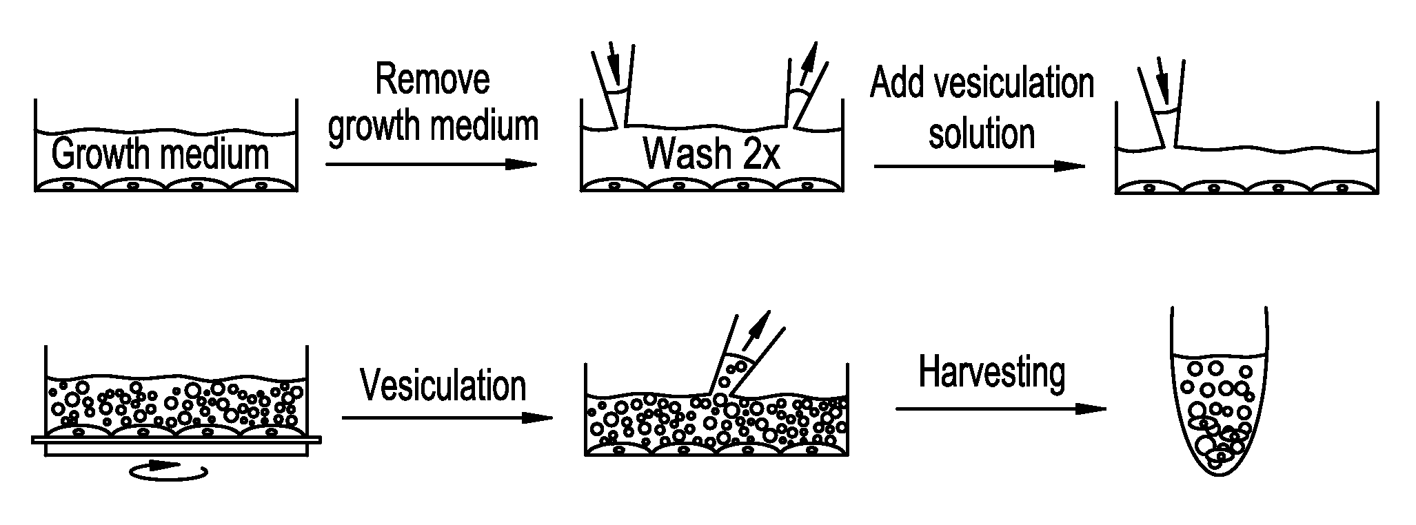
[0159]The production of high-purity plasma membrane vesicles consists of four steps:
[0160]1. Formation of PMVs by addition of PMV-forming agents to a cell culture
[0161]2. Release of PMVs from cell culture
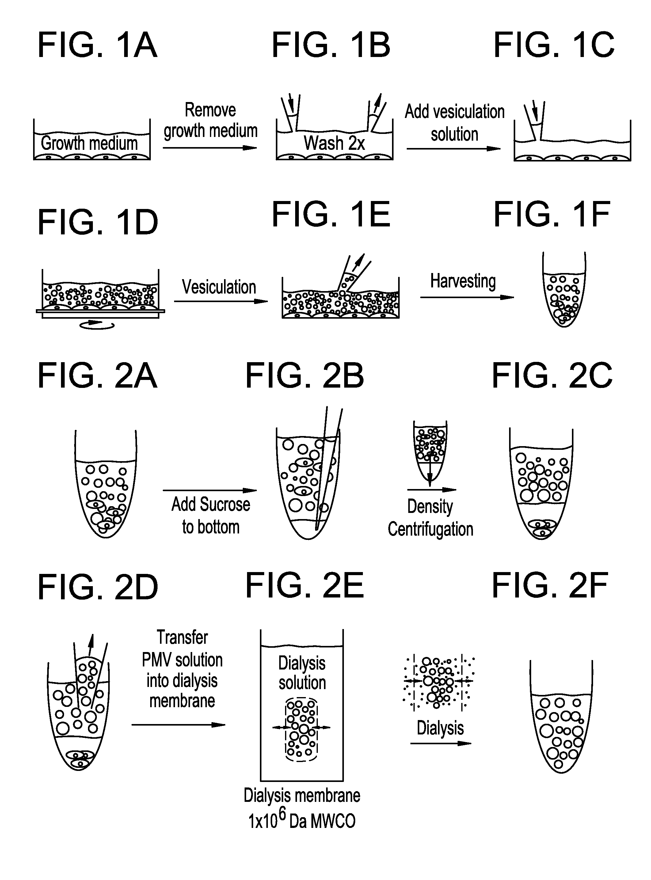
[0162]3. Purification of PMVs by density gradient centrifugation
[0163]4. Purification of PMVs by dialysis
[0164]Steps 1-2 yields a crude polydisperse PMV fraction with sizes of PMVs ranging up to about ten micrometers in diameter. Such PMVs can find great use in several structural and functional assays. Examples of such assays include ion channel function, G-Protein function, adhesion protein function, and many more.
[0165]Adding steps 3 and 4 yield a cell-free ultrapure polydisperse PMV fraction with sizes of PMVs ranging up to about ten micrometers in diameter that can be utilized in several structural and functional assays, including proteomic assays to screen for protein expression, and target identification.
[0166]Formation ...
example 2
Isolation and Purification of Plasma Membrane Vesicles from NG-108 Cells and Subsequent LC-MS / MS Proteomic Analysis
[0196]Isolation and purification of plasma membrane vesicles. NG108-15 cells were grown to confluence using DMEM (4,5 g / L glucose, 2mM L-glutamine) with 10% FCS. Vesiculation was performed as previously described20,29 with some modifications. Briefly, the confluent cell layer was washed twice with 10 mM HEPES, 140 mM NaCl, pH 7.4. Cells were incubated with 2-4 mL vesiculation buffer (10 mM Hepes, 140 mM NaCl containing 2 mM CaCl2, 2 mM DTT, and 25 mM formaldehyde, pH 7.4), where the incubation time was chosen between 8-16 hours at 37° C., with gentle shaking (60-80 cycles / minute). The supernatant was collected from the flasks and pooled in a 15 mL conical tube. In order to remove cells that have detached from the surface, the solution was underlaid with 2 mL 2M sucrose and centrifuged 15 min at 4° C. with 500×g using a swing-out rotor. The supernatant was collected, and...
example 3
Extraction of Lipid Components
[0207]CHO-K1 cells were cultured to 95% confluency in T175 flasks. Media was removed from the flasks, and the cells were washed several times with 150 mM NaCl, 10 mM HEPES, 2 mM CaCl2, pH 7.4. To induce plasma membrane vesiculation, 6 mL of a solution of 25 mM formaldehyde, 2 mM DTT, 150 mM NaCl, 10 mM HEPES, 2 mM CaCl2, pH 7.4, was added to each flask. Vesiculation was done for 2 hours at 37 C, with gentle rocking of the cell flasks. After vesiculation, the solutions containing plasma membrane vesicles were collected from the flasks and pooled. The solution was passed through a 40 μm pore filter to remove aggregates of cells, and through a 5 μm filter to remove single cells. The solution was then frozen at −20C. The blebb solutions from different cell batches were pooled into larger batches for extraction. The total blebb solution volume was measured and used to calculate the organic solvent volumes for extraction. The first step was to add NH4Ac (ammo...
PUM
| Property | Measurement | Unit |
|---|---|---|
| pH | aaaaa | aaaaa |
| diameter | aaaaa | aaaaa |
| diameter | aaaaa | aaaaa |
Abstract
Description
Claims
Application Information
 - R&D Engineer
- R&D Manager
- IP Professional
- Industry Leading Data Capabilities
- Powerful AI technology
- Patent DNA Extraction
Browse by: Latest US Patents, China's latest patents, Technical Efficacy Thesaurus, Application Domain, Technology Topic.
© 2024 PatSnap. All rights reserved.Legal|Privacy policy|Modern Slavery Act Transparency Statement|Sitemap