Identification of high grade or >= cin2 for early stages and late stages detection, screening, and diagnosis of human papillomavirus (HPV) and hpv-associated cancers

Active Publication Date: 2012-03-01
HEER MEDICAL TECH DEV CO LTD
View PDF6 Cites 2 Cited by
  • Summary
  • Abstract
  • Description
  • Claims
  • Application Information

AI Technical Summary

Benefits of technology

[0039]In one embodiment, the papillomavirus comprises one or more viruses selected from the group consisting of HPV-6, HPV-11, HPV-16, HPV-18, HPV-31, HPV-33, HPV-35, HPV-39, HPV-42, HPV-43, HPV-44, HPV-45,

Problems solved by technology

The test is subjective with significant inter-observer variability, and is limited by low sensitivity and high false positive results.
Although colposcopic biopsy has historically been considered the gold standard, recent reports indicate that cervical biopsies may miss 33% to 50% of high-grade disease because of sampling or diagnostic errors.
As a result, it may be difficult to differentiate between false positive cervical cytology results versus false-negative biopsy results.
The presence of these lesions, preferentially observed in women aged 35-40 yrs, are at high risk of progression toward invasive cervical cancer.
However, due to subjective test criteria, drawbacks of Pap smear tests include difficulty in obtaining samples, poor inter- and intra-observer agreement, a high rate of false negatives (up to 20%) and false positive, the requirements for specialized laboratories staffed with highly trained personnel, and inability to identify a large proportion of HPV-infected persons.
Detecting HPV infection by nucleic acid methods, has been developed, but not ideal, due to not only its high cost, assay operation procedures, the requirements for facility, equipment, and highly trained personnel, but also its very low positive predictive value to CIN.
In addition, DNA testing could not differentiate the diagnosis of LSIL from HSIL, nor CIN lesions from non-transforming latent or remissive viral infection.
However, there are limited reports showing direct detection

Method used


Image

  • Identification of high grade or >= cin2 for early stages and late stages detection, screening, and diagnosis of human papillomavirus (HPV) and hpv-associated cancers
  • Identification of high grade or >= cin2 for early stages and late stages detection, screening, and diagnosis of human papillomavirus (HPV) and hpv-associated cancers
  • Identification of high grade or >= cin2 for early stages and late stages detection, screening, and diagnosis of human papillomavirus (HPV) and hpv-associated cancers

Examples


example 1

Detailed Example 1

[0097]Some embodiments are directed to monoclonal antibodies against HPV proteins. Obtaining high quantity of purified recombinant HPV proteins in native conformation as immunogen becomes the first critical step in generating antibodies specific for detecting HPV proteins in clinical samples. E6 and E7 is known to be very difficult to isolate and purify due to undesirable aggregation during protein purification, protein instability, low levels of expression, and low immunogenic responses of purified proteins. We have overcome the technical barriers to produce HPV E6 and E7 recombinant protein in a non-denatured, soluble form. To demonstrate that the purification method that we have developed results in a conformation closely approximating the native form to bind the anti-HPV antibodies, we have used the HPV infected cervical samples (high risk-HPV positive by PCR) that contain human HPV antibody to test the purified recombinant HPV proteins. Studies of using such p...

example 2

Detailed Example 2

Immunocytochemistry Assay (ICC)

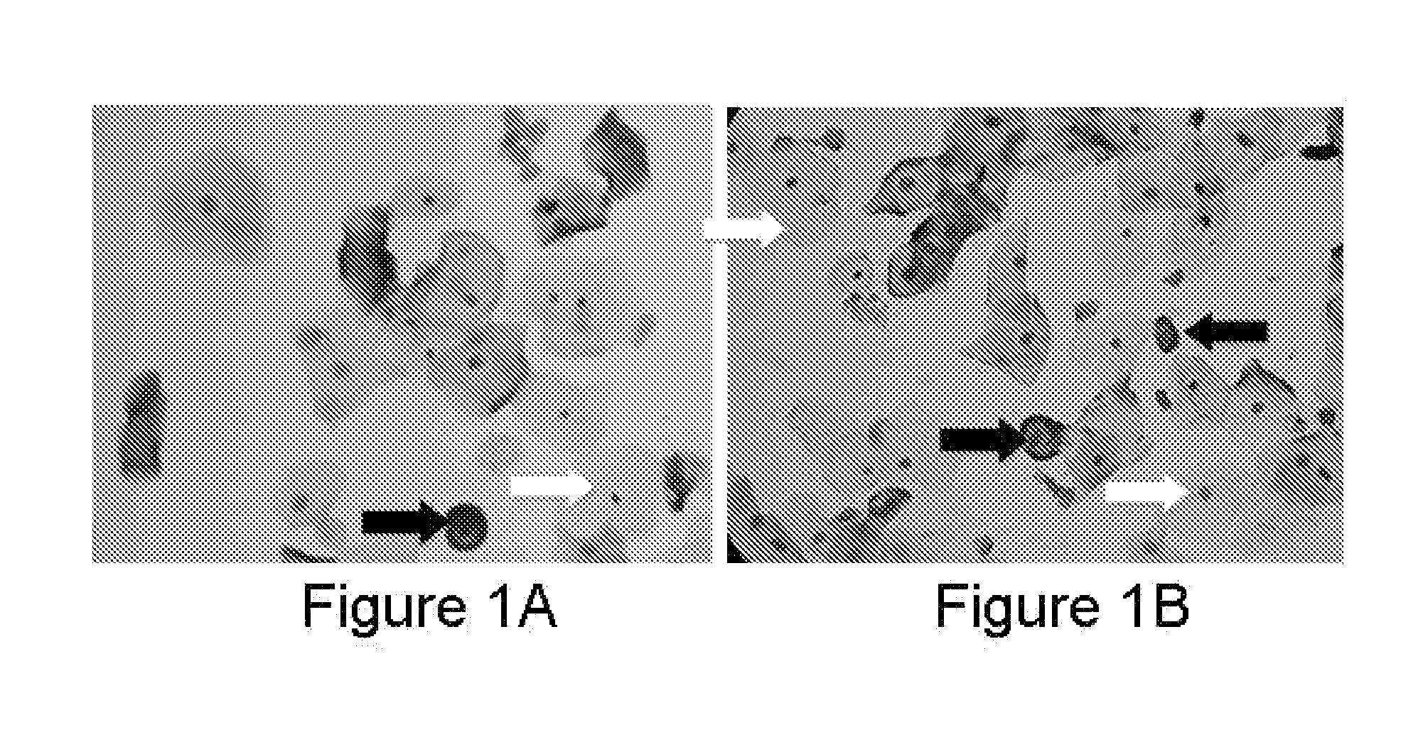
[0103]Some embodiments are directed to immunoassay comprising the detection of HPV proteins and cellular proteins in exfoliated cervical cells. Sample preparation: The cervical scrape cells collected from liquid based solution were divided into two parts, one for cytological Papanicolaou staining, and another one for immunocytochemical staining using HPV antibodies described in some embodiments. In the Pap smear results, Papanicolaou staining samples were scored 0-17. Score one (1) to three (3) are considered as normal, and score four (4) and above as abnormal. Interpretation of Pap smear reports can be challenging at times. Based on the Bethesda 2001 system, negative Pap smear may include negative intraepithelial lesion or malignancy, and abnormal Pap smear may include different stages of squamous cells in development of dysplasia or lesions. For examples, LSIL: Low grade of Squamous Intraepithelial Lesion, HSIL: High grade of Squamo...

example 3

Detailed Example 3

Immunohistochemistry Assay (IHC)

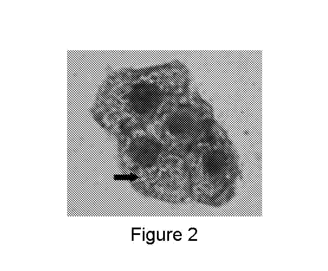
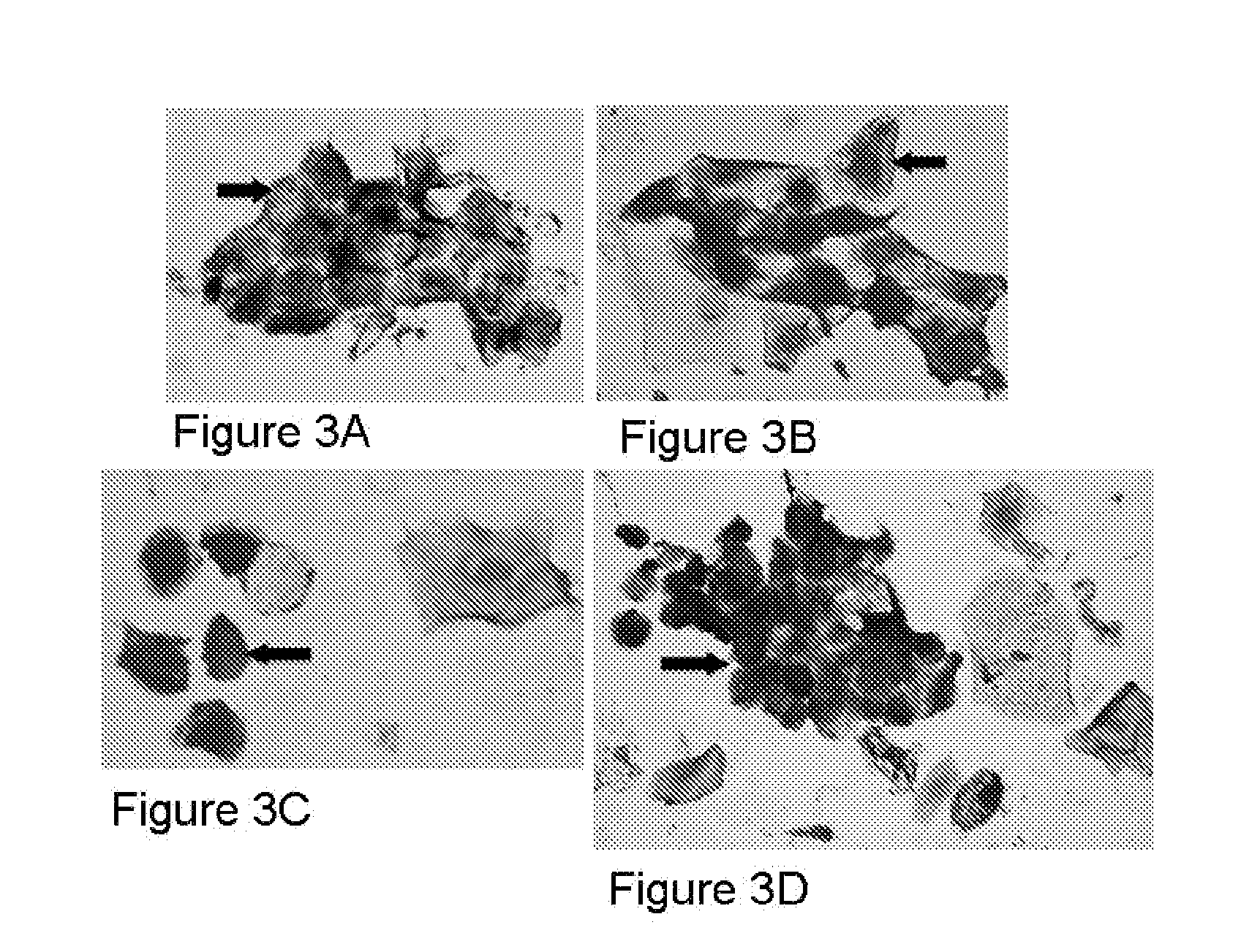
[0135]Some embodiments are directed to a method of screening a human subject of Papillomavirus infection includes providing a thin section containing one or more kinds of tissue cells from a clinical tissue sample of the human subject, applying the thin section on a slide, conducting one or more immunohistochemical assays on the slide containing the thin section of the clinical tissue sample, staining the thin layer of human cells using one or more antibodies generated against one or more purified recombinant Papillomavirus proteins, wherein at least one antibody is capable of recognizing a Papillomavirus early protein, and detecting in situ one or more proteins from one or more Papillomavirus types present in the thin section of the clinical tissue sample on the slide.

[0136]Sample preparation: Paraffin tissues blocks sectioned into 4 microns were placed on slide and baked at 60° C. overnight. Deparaffin / hydrate sections were unmaske...


PUM


Abstract

A method and a kit for determining a disease stage of a papilloma virus infection in a human subject is disclosed. The method comprises: obtaining a sample from said human subject, said sample consisting of a tissue sample comprising proteins or a cell sample comprising proteins; contacting said sample with one or more antibodies that specifically bind to one or more recombinant HPV proteins; determining a presence, absence, or amount of at least one protein in said sample that specifically binds to said antibodies and determining said disease stage of papilloma virus infection in said human subject based on said determined presence, absence, or amount of said one or more proteins.

Description

CROSS-REFERENCE TO RELATED APPLICATIONS[0001]This application claims the benefit of U.S. Prov. Patent App. Ser. No. 61 / 215,589, filed on May 7, 2009, entitled “Novel Antibodies and Assays Useful for Early Stages and Late Stages Detection, Screening, and Diagnosis of Human Papillomavirus (HPV) and HPV-Associated Cancers”.[0002]This application is cross related to U.S. patent application Ser. No. 11 / 559,366, filed on Nov. 13, 2006, entitled “Detection Method for Human Papillomavirus (HPV) and Its Application in Cervical Cancer”, U.S. patent application Ser. No. 12 / 082,740, filed on Apr. 14, 2008, entitled “Protein Chips for HPV Detection”, U.S. App. Ser. No. 61 / 131,991, filed on Jun. 13, 2008, entitled “Antibodies and Assays for HPV Detection”, and U.S. App. Ser. No. 61 / 192,912, filed on Sep. 22, 2008, entitled “Novel Monoclonal Antibodies against HPV Proteins Useful for Early State and Late Stage Detection, Screening, and Diagnosis of HPV Related Cervical Cancer”, U.S. application Se...

Claims


Application Information

Patent Timeline
IPC IPC(8): C12Q1/70
CPCG01N33/56983G01N2800/56G01N2333/025G01N33/57411
Inventor CHENG, SHULING
Owner HEER MEDICAL TECH DEV CO LTD
Why Eureka
Try Eureka