Radiographic image obtainment method and radiographic apparatus

a radiographic apparatus and obtainment method technology, applied in the direction of material analysis using wave/particle radiation, applications, instruments, etc., can solve the problems of inability to obtain sufficient contrast in an image, inability to obtain x-ray transmission images with sufficient intensity (contrast), and small difference in x-ray absorption between the two, so as to reduce the influence of moiré, reduce the position shift, and achieve higher quality radiographic images

Inactive Publication Date: 2012-06-28
FUJIFILM CORP
View PDF0 Cites 31 Cited by
  • Summary
  • Abstract
  • Description
  • Claims
  • Application Information

AI Technical Summary

Benefits of technology

[0062]According to a radiographic image obtainment method and a radiographic apparatus of the present invention, the first and second gratings and/or the radiation image detector is structured in such a manner that they are attachable to the radiographic apparatus and detachable therefrom. Further, when attachment or detachment of the first and second gratings and/or the radiation image detector is detected, a radiation source is controlled so as to perform preliminary irradiation to detect a relative positional deviation (shift in position, misregistration, misalignment, displacement or the like) between the first and second gratings and the radiation image detector. Therefore, for example, if a position shift is detected based on an image for checking

Problems solved by technology

Since a difference in X-ray absorptivity is small in soft tissue of a living body, soft material, and the like, a sufficient difference in intensity (contrast) as an X-ray transmission image is not obtainable.
Therefore, a difference in X-ray absorptivity between the two is small, and a sufficient contrast in an image is hard to obtain.
However, imaging by MRI has problems of a long imaging time of several tens of minutes, a low image resolution of about 1 mm, and a low cost-effectiveness which is difficult to be adopted in regular checkups of physical examination.
However, such a facility is too large to be adopted in general hospitals.
Therefore, it is extremely difficult to directly measure the shift in the position.
However, since the phase shift amount of moiré fringes is also very small, a small fluctuation of the moiré image greatly affects the phase restoration accuracy.
Further, since a cassette attaching/detaching mechanism

Method used


Image

  • Radiographic image obtainment method and radiographic apparatus
  • Radiographic image obtainment method and radiographic apparatus
  • Radiographic image obtainment method and radiographic apparatus

Examples


Embodiment Construction

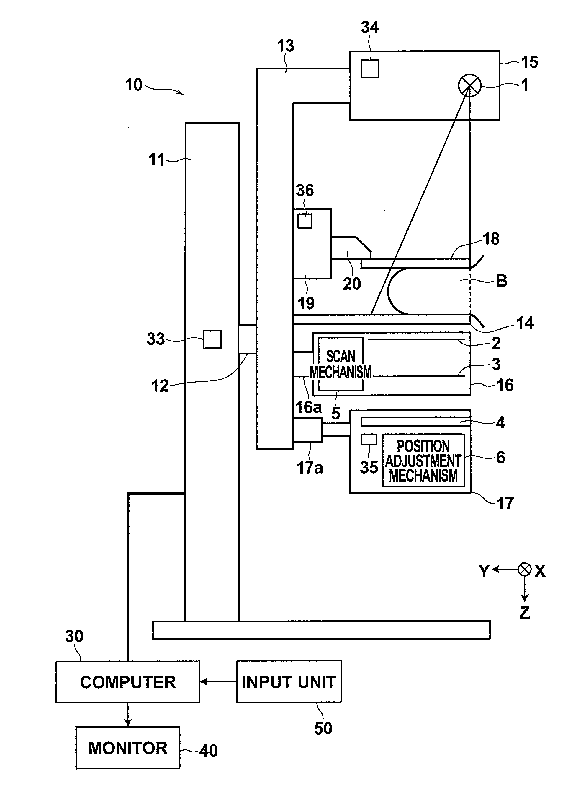
[0107]Hereinafter, a mammography and display system using an embodiment of a radiographic apparatus according to the present invention will be described with reference to drawings. FIG. 1 is a schematic diagram illustrating the configuration of the whole mammography and display system using an embodiment of the present invention.

[0108]As illustrated in FIG. 1, the mammography and display system of the present invention includes a mammography apparatus 10, a computer 30 connected to the mammography apparatus 10, a monitor 40 and an input unit 50. The monitor 40 and the input unit 50 are connected to the computer 30.

[0109]As illustrated in FIG. 1, the mammography apparatus 10 includes a base 11, a rotation shaft 12, and an arm 13. The rotation shaft 12 is movable in a vertical direction (Z direction) with respect to the base 11, and rotatable. The arm 13 is connected to the base 11 by the rotation shaft 12.

[0110]The arm 13 is alphabet “C” shaped. A radiography table 14 on which breast...


PUM


Abstract

In a radiographic apparatus, a radiation image detector or first and second gratings are structured in such a manner to be attachable to the radiographic apparatus and detachable therefrom. The radiographic apparatus includes a cassette attachment/detachment detection unit that detects attachment and detachment of the radiation image detector, or a grid attachment/detachment detection unit that detects attachment and detachment of the first and second gratings. The apparatus further includes a preliminary irradiation control unit that controls a radiation source so that preliminary irradiation for detecting a relative positional deviation between the first and second gratings and the radiation image detector is performed when attachment or detachment of the radiation image detector, or the first and second gratings has been detected.

Description

BACKGROUND OF THE INVENTION[0001]1. Field of the Invention[0002]The present invention relates to a radiographic image obtainment method and a radiographic apparatus using a grating or gratings.[0003]2. Description of the Related Art[0004]Since X-rays attenuate depending on the atomic number of an element constituting a substance through which the X-rays pass, and the density and the thickness of the substance, the X-rays are used as a probe for observing the inside of a subject from the outside of the subject. X-ray radiography is widely used in medical diagnosis, non-destructive examination, and the like.[0005]In a general X-ray radiography system, a subject is placed between an X-ray source for emitting X-rays and an X-ray image detector for detecting an X-ray image. In this state, radiography is performed on the subject to obtain a transmission image of the subject. In this case, each X-ray emitted from the X-ray source toward the X-ray image detector attenuates (is absorbed) by ...

Claims


Application Information

Patent Timeline
IPC IPC(8): G01N23/04
CPCA61B6/4283A61B6/484A61B6/502G01T1/247A61B6/4092A61B6/4291G01T1/24G01T1/246
Inventor IWAKIRI, NAOTOISHII, HIROYASU
Owner FUJIFILM CORP
Why Eureka
Try Eureka