In other cases, the pathology may be minor at the outset but, if left untreated, may worsen over time.
The pathology may, either initially or thereafter, significantly interfere with patient comfort and lifestyle.
In some cases, the pathology can be so severe as to require partial or total hip replacement.
A number of procedures have been developed for treating hip pathologies short of partial or total hip replacement, but these procedures are generally limited in scope due to the significant difficulties associated with treating the hip joint.
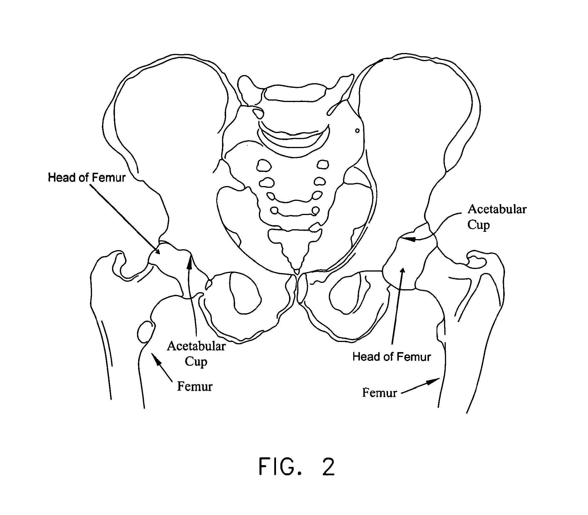
In some cases, and looking now at FIG. 13, this impingement can occur due to irregularities in the geometry of the femur.
In other cases, and looking now at FIG. 14, the impingement can occur due to irregularities in the geometry of the acetabular cup.
Impingement can result in a reduced range of motion, substantial pain and, in some cases, significant deterioration of the hip joint.
Defects of this type sometimes start out fairly small but often increase in size over time, generally due to the dynamic nature of the hip joint and also due to the weight-bearing nature of the hip joint.
Articular defects can result in substantial pain, induce and / or exacerbate arthritic conditions and, in some cases, cause significant deterioration of the hip joint.
More particularly, in many cases, an accident or sports-related injury can result in the labrum being torn away from the rim of the acetabular cup, typically with a tear running through the body of the labrum.
These types of injuries can be very painful for the patient and, if left untreated, can lead to substantial deterioration of the hip joint.
Unfortunately, minimally-invasive treatments for pathologies of the hip joint have lagged far behind minimally-invasive treatments for pathologies of the shoulder joint and the knee joint.
As a result, it is relatively difficult for surgeons to perform minimally-invasive procedures on the hip joint.
This limited access further complicates effectively performing minimally-invasive procedures on the hip joint.
In addition to the foregoing, the nature and location of the pathologies of the hip joint also complicate performing minimally-invasive procedures on the hip joint.
This makes drilling into bone, for example, significantly more complicated than where the angle of approach is effectively aligned with the angle at which the instrument addresses the tissue, such as is frequently the case in the shoulder joint.
Furthermore, the working space within the hip joint is typically extremely limited, further complicating repairs where the angle of approach is not aligned with the angle at which the instrument addresses the tissue.
As a result of the foregoing, minimally-invasive hip joint procedures are still relatively difficult to perform and relatively uncommon in practice.
Consequently, patients are typically forced to manage their hip pain for as long as possible, until a resurfacing procedure or a partial or total hip replacement procedure can no longer be avoided.
These procedures are generally then performed as a highly-invasive, open procedure, with all of the disadvantages associated with highly-invasive, open procedures.
However, due to the anatomy of the hip joint and the pathologies associated with the same, hip arthroscopy is currently practical for only selected pathologies and, even then, hip arthroscopy has generally met with limited success.
Unfortunately, current methods and apparatus for arthroscopically repairing (e.g., re-attaching) the labrum are somewhat problematic.
Unfortunately, bone anchors of the sort described above are traditionally used for re-attaching ligaments to bone and, as a result, tend to be relatively large, since they must carry the substantial pull-out forces normally associated with ligament reconstruction.
However, this large anchor size is generally unnecessary for labrum re-attachment, since the labrum is not subjected to substantial pull-out forces, and the large anchor size typically causes unnecessary trauma to the patient.
Furthermore, the large size of traditional bone anchors can be problematic when the anchors are used for labrum re-attachment, since the bone anchors generally require a substantial bone mass for secure anchoring, and such a large bone mass is generally available only a substantial distance up the acetabular shelf.
However, labral re-attachment utilizing a bone anchor set high up into the acetabular shelf creates a suture path, and hence a labral draw force, which is not directly aligned with the portion of the acetabular rim where the labrum is to be re-attached.
This can sometimes result in a problematic labral re-attachment and, ultimately, can lead to a loss of the suction seal between the labrum and femoral head, which is a desired outcome of the labral re-attachment procedure.
Alternatively, the suture path can also surround the labrum, thus placing a suture on both sides of the labrum, including the articular side of the labrum, and thus exposing the articular surface of the femur to a foreign body, which could in turn cause damage to the articular surface (i.e., the articular cartilage) of the femur.


