Method of detecting rupture of membranes

Inactive Publication Date: 2013-05-30
BIOSYNEX
View PDF4 Cites 3 Cited by
  • Summary
  • Abstract
  • Description
  • Claims
  • Application Information

AI Technical Summary

Benefits of technology

[0108]Embodiments of the present invention make it possible to detect ruptures of fetal membranes whatever their size, for example whether they are microruptures and/or macroruptures, and/or whatever the volume of amniotic fluid sampled.
[0109]Embodiments of the present invention make it possible to reduce the probability of obtaining false-negative results and also false-positive results.
[0110]In add

Problems solved by technology

During pregnancy, women may be subjected to various factors and/or events that can endanger the pregnancy.
The rupture of fetal membranes is one of these events, particularly when the rupture of the membranes is premature.
Moreover, the loss of amniotic fluid leads to a decrease in the space available for the development of the fetus.
This decrease can, when the rupture takes place before the period of 24 weeks of amenorrhea, endanger the vital prognosis of the fetus, leading to a miscarriage and/or resulting in the birth of a non-viable baby.
After this 24-week period, the vital prognosis of the infant is still in question, resulting in premature labor and/or an infection of the fetus that may result in voluntary inducing of labor.
Moreover, the decrease in the volume of amniotic fluid can cause problems of malformation, limb atrophy, and developmental abnormality of various parts of the body.
This diagnostic test is a test which is not very sensitive and which is not very suitable for the diagnosis of microruptures, which does not make it possible to rapidly and easily detect the rupture of membranes, and which is especially related to the ability of the practitioner to be able to identify/visualize the flow of the fluid.
However, these tests are not reliable and can lead to false-negative re

Method used


Image

  • Method of detecting rupture of membranes

Examples


Example

Example 1

Method of Detecting Rupture of Fetal Membranes by Means of an Immunofiltration Device

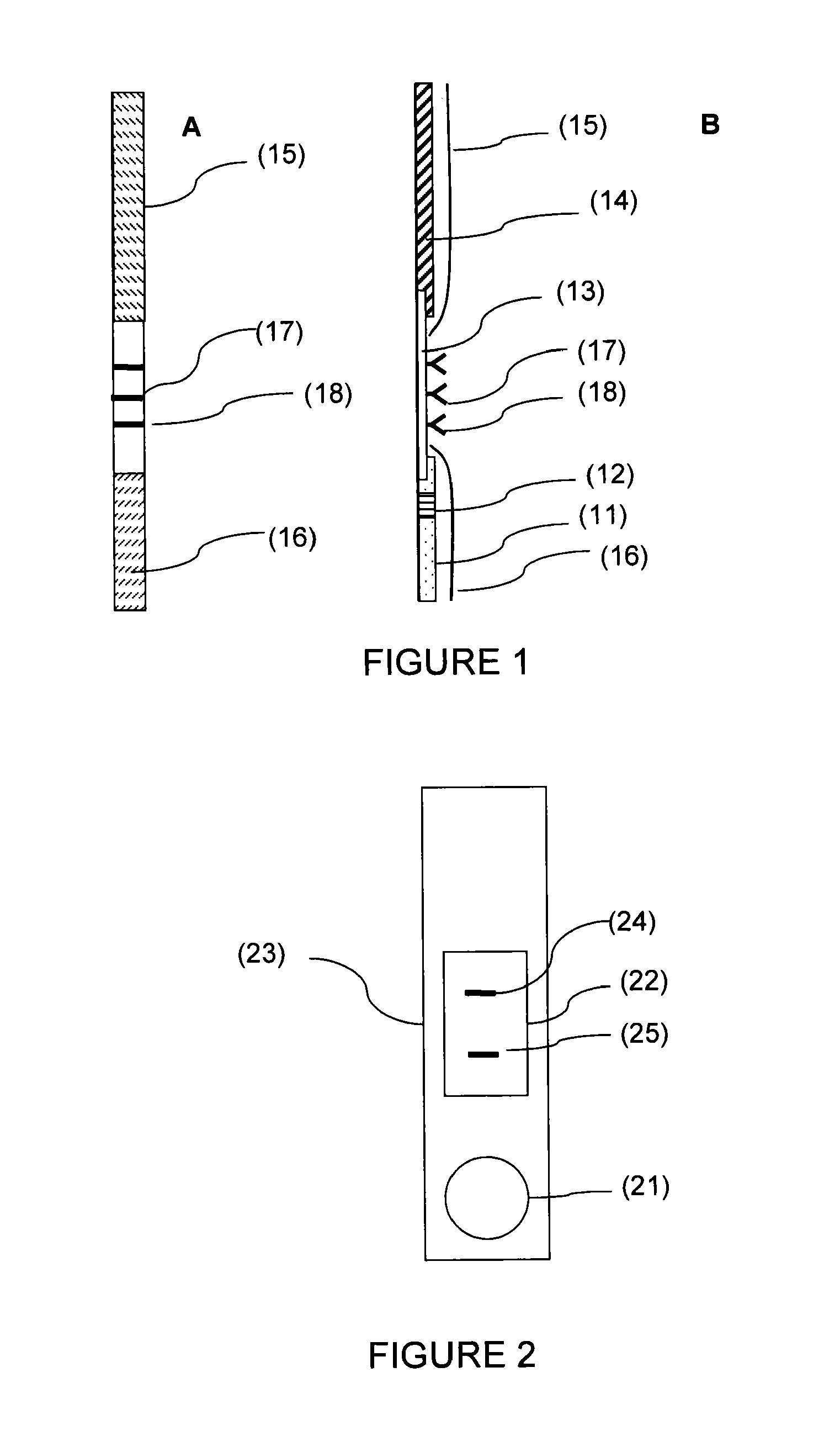
[0123]Anti-AFP antibodies (ABAFP-0404, clone 4, Arista Biologicals) diluted to 0.75 mg / ml in a PBS buffer and also anti-IGFBP-1 antibodies (I 2032, clone 33627.11, Sigma Aldrich) diluted to 0.75 mg / ml in a PBS buffer are prepared.

[0124]The antibodies are deposited, using a reagent-distributing automated device (Isoflow Dispenser, Imagene Technology Inc. (registered trademark)), in the form of parallel lines on a nitrocellulose membrane, and then dried in an incubator at 37° C. under an atmosphere with a controlled degree of humidity of less than 30%. The membrane is then cut up into multiple strips approximately 1 centimeter wide and 2.5 centimeters long. Each strip is then placed on an absorbent support placed in a plastic base. A plastic cover which fits the base is then placed over the base and closed by manual pressure. The plastic cover has a window opening, the surface area of which i...

Example

Example 2

Immunochromatographic Device for Detecting Rupture of Fetal Membranes

[0128]The strips are prepared from laminated cards which are 30 cm×9.5 cm (CNPC-SS12 R-032 / 2, MDI (registered trademark)) consisting of a plastic support covered with an adhesive layer on which the nitrocellulose membrane, the absorbent paper and the glass fiber are assembled.

[0129]Using a reagent-distributing automated device (Isoflow Dispenser, Imagene Technology Inc. (registered trademark)), goat anti-mouse IgG antibodies (ABGAM-0500, Arista Biologicals (registered trademark)) diluted to 4 mg / ml in a PBS buffer, anti-AFP antibodies (ABAFP-0404, clone 4, Arista Biologicals) diluted to 0.75 mg / ml in a PBS buffer and anti-IGFBP-1 antibodies (I 2032, clone 33627.11, Sigma Aldrich (registered trademark)) diluted to 0.75 mg / ml in a PBS buffer are deposited with a flow rate of 0.5 μl / cm in the form of parallel lines 5 mm apart and each from 1 to 2 mm wide, on a nitrocellulose membrane which is 25 mm wide (zone...

Example

Example 3

Example of Use of a Device for Implementing the Method of the Invention

[0139]1. A solution of 2.5 ml of 50 mM borate (02102391, Biosolve), 1% BSA (AP-4510-01, Seracare Life Sciences) and 0.05% triton X-100 (pH 9.3) was introduced into a 5 ml screw-cap tube (015610, Dustcher),[0140]2. the sample was taken with a sterile swab having a polyester bud and deposited on the device described in example 2,[0141]3. the device obtained was introduced into the abovementioned screw-cap tube,[0142]4. the result is obtained after 10 minutes of migration.[0143]The test is positive when at least one of the two bands corresponding to AFP and to IGBPF-1 is colored.

[0144]When the test is positive, the veracity of the test can be verified by a complete gynecological examination making it possible to confirm or refute the result.


PUM


Abstract

The present invention relates to a method of detecting rupture of fetal membranes and to a device for implementing said method. In particular, a method of in-vitro detection of a rupture of fetal membranes comprising a step of simultaneously searching, within a specimen of vaginal or cervical secretions, for the alpha fetal protein (AFP) and for the insulinomimetic growth factor-binding protein 1 (IGFBP-1). The present invention finds, in particular, an application in the medical field, especially in the obstetric field.

Description

PRIORITY CLAIM[0001]This application is a National Phase entry of PCT Application No. PCT / FR2011 / 051254, filed Jun. 1, 2011, which claims priority from French Application No. 1054374 filed Jun. 3, 2010, the disclosures of which are hereby incorporated by referenced herein in their entirety.TECHNICAL FIELD[0002]The present invention relates to a method of detecting rupture of fetal membranes and to a device for implementing said method.[0003]The present invention finds in particular an application in the medical field, the diagnostic field, in particular in the obstetric field.[0004]In the description below, the references between square brackets ([ ]) refer to the list of references provided at the end of the text.BACKGROUND OF THE INVENTION[0005]During pregnancy, women may be subjected to various factors and / or events that can endanger the pregnancy. The rupture of fetal membranes is one of these events, particularly when the rupture of the membranes is premature.[0006]The origin o...

Claims


Application Information

Patent Timeline
IPC IPC(8): G01N33/68
CPCG01N33/689G01N2800/368G01N2333/4745G01N2333/471
Inventor PAPER, THIERRYRAKOTONDRAMANGA, FRANCOISE
Owner BIOSYNEX
Why Eureka
Try Eureka