

Multilayered silk scaffolds for meniscus tissue engineering
a meniscus and tissue engineering technology, applied in the field of meniscus tissue engineering scaffolds, can solve the problems of predisposing patients to osteoarthritis and affecting normal knee function, so as to improve outcomes, prevent patients from osteoarthritis, and improve knee function
- Summary
- Abstract
- Description
- Claims
- Application Information
AI Technical Summary
Benefits of technology
Problems solved by technology
Method used
Image

Examples
example 1
Gross Morphology and Stacking of Engineered Silk Meniscal Constructs
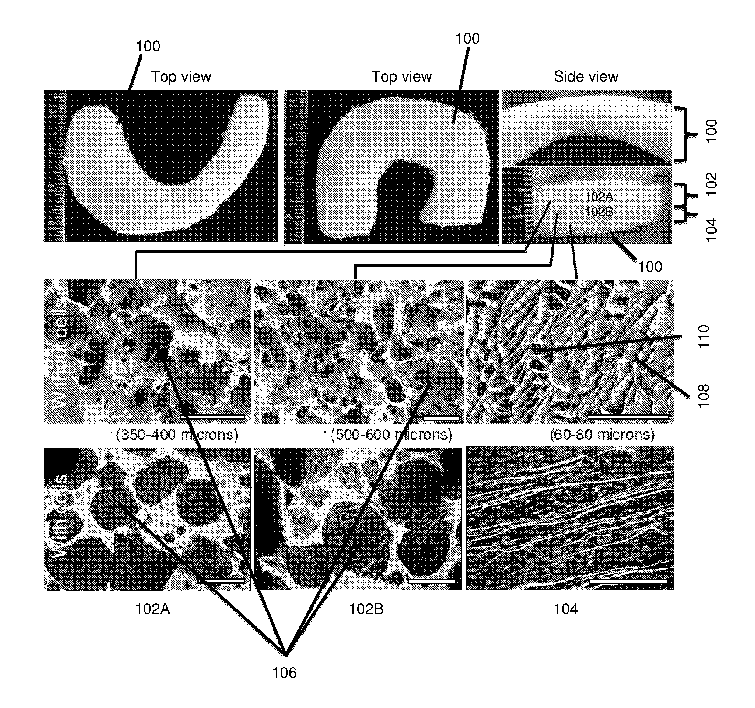
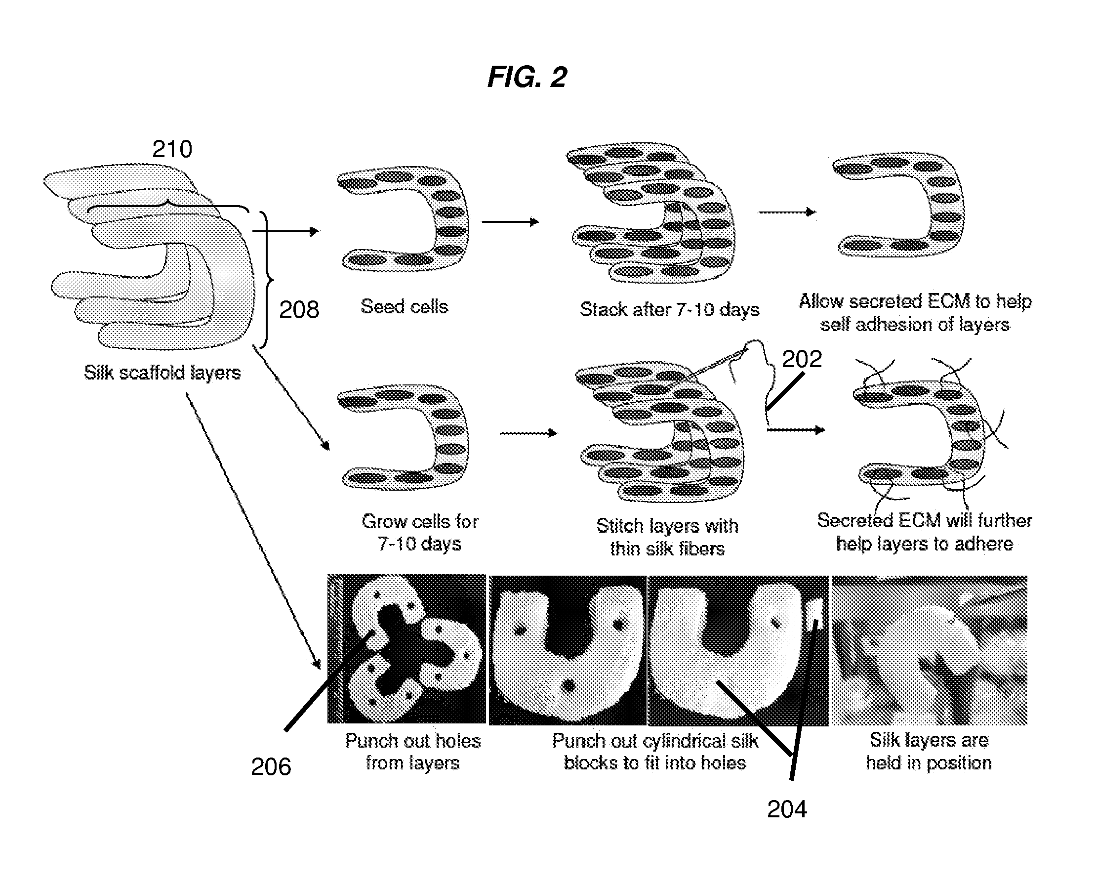
[0229]Native meniscus pore heterogeneity was mimicked within silk scaffolds as individual layers using an all aqueous method (FIG. 1). In some embodiments, the fabricated scaffold layers can be rigid and compact due to relatively high (9 wt %) silk content. Scanning electron microscopic (SEM) pictures revealed that the top two layers fabricated using salt leaching method had circular pores in the size range of about 350-about 400 and about 500-about 600 microns, respectively. The pores were highly interconnected with smaller pores. However, the bottom third layer fabricated using freeze-drying method showed laminar channels with pore width ranging between 60-80 microns. The fabricated scaffolds were shaped like native meniscus (wedge shaped) and three individual layers were stacked on top of each other to form a single unit mimicking meniscus multiporosity (FIG. 1). FIG. 2 demonstrates exemplary methods that can be ...
example 2
Cell Attachment and Proliferation on Individual Silk Scaffold Layers
[0230]Confocal microscopy was used to evaluate fibroblast and chondrocyte cell proliferation and attachment onto individual silk meniscus scaffold layers. At day 1, cells appeared clustered and attached onto walls of scaffolds in their early stage of seeding. Further, cell spreading was observed to be minimal at day 1 in both cases (fibroblasts and chondrocytes) as the scaffold pore lumens can be clearly seen (FIGS. 3A to 3I, 4A to 4C). In comparison, after day 28 of seeding and culture, the cells had filled the scaffold pore voids and spread out actin filaments. In all three meniscal scaffold layers the cells (fibroblast and chondrocytes) were evenly distributed and showed confluence (FIGS. 3A to 3I, 4D to 4F). Further, PicoGreen DNA assay results showed that the chondrocytes proliferated with an increase of approximately ˜48%, ˜64% and ˜38% compared to initial cell numbers (day 28 vs. day 1) in the case of 1st, 2n...
example 3
Histology and Immunocytochemistry of Individual Meniscus Scaffold Layers
[0231]Histology sections of individual meniscus scaffold layers revealed accumulation of sGAG and collagen with time (day 28 vs day 1) in all three layers. Compared to day 1, samples for day 28 stained deeper and higher for Alician blue, Saffranin O and H & E in all three layers for both cell types (FIGS. 5A to 5C, 6A to 6C). For Alician blue, chondrocytes showed intense blue color indicating presence of abundant sGAG. Particularly for the laminar 3rd scaffold layer, the cells stained intense deep blue indicating matured chondrocyte phenotype (FIGS. 6B, 8). For fibroblast, the staining was lighter compared to chondrocytes, but showed positive blue color on culturing in chondrogenic medium (FIGS. 5B, 8). Similarly, Saffranin O staining confirmed GAG accumulation within scaffold pores in all layers. In case of Saffranin O staining, faint positive staining of fibroblast cells was observed compared to intense staini...
PUM
| Property | Measurement | Unit |
|---|---|---|
| Length | aaaaa | aaaaa |
| Length | aaaaa | aaaaa |
| Length | aaaaa | aaaaa |
Abstract
Description
Claims
Application Information