Biomarker of liver cancer and uses thereof
a liver cancer and biomarker technology, applied in the field of liver cancer biomarkers and nucleolin-based methods for detecting or treating liver cancer, can solve the problems of insufficient monitoring of liver cancer, many new molecular markers of liver cancer have not been discovered, and complicated liver cancer
- Summary
- Abstract
- Description
- Claims
- Application Information
AI Technical Summary
Problems solved by technology
Method used
Image

Examples
example 1
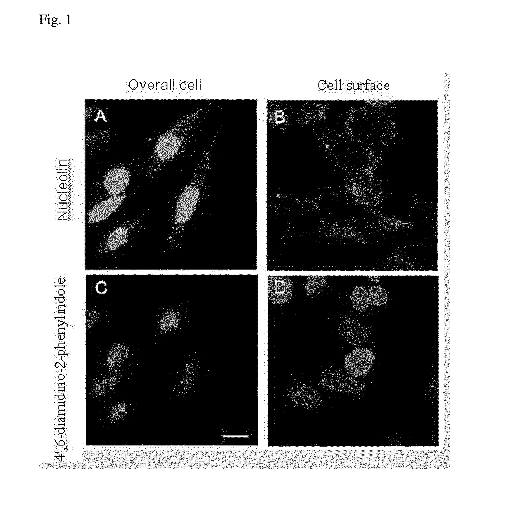
Distribution of Nucleolin Protein in Liver Cells
[0055]Liver cancer cells SK-Hep1 were washed by phosphate buffer solution three times, and then 4% paraformadehyde was added as a fixative. All cells were covered by the fixative and allowed to stand at 37° C. for 10 minutes. The fixative was removed, followed by addition of phosphate buffer solution. After slowly shaken at room temperature for 1 hour, the cells were washed by phosphate buffer solution three times. The cells to be holed were soaked in Triton X-100 solution with concentration of 2% for 10 minutes, washed by phosphate buffer solution three times, and then 1% bovine serum albumin was used to remove non-specific binding. After slowly shaken at room temperature for 1 hour to absorb 1% bovine serum albumin, the cells were washed by phosphate buffer solution three times. Nucleolin primary antibody (purchased from Santa Cruz Biotechnology, Santa Cruz, Calif.; Catalog No.: sc-8031) diluted 200 times was added, slowly shaken at ...
example 2
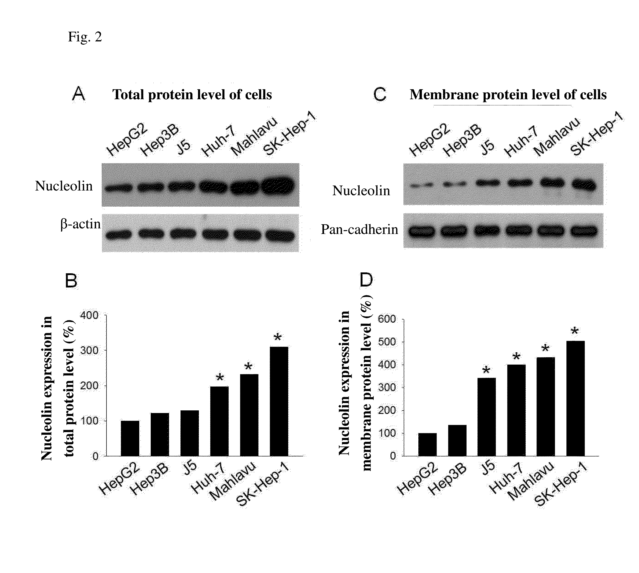
Nucleolin Protein Expression Level in Different Liver Cancer Cell Lines
[0058]The human liver cancer cell lines HepG2 and Hep3B used in the example were cultured in MEM medium supplemented with 10% fetal bovine serum (FBS, purchased from Invitrogen Carlsbad, USA). Mahlavu, J5, Huh-7, and SK-Hep-1 cells were cultured in DMEM medium supplemented with 10% FBS. All mediums were additionally added 100 U / ml penicillin and 100 μg / ml streptomycin and were incubated at 37° C. with carbon dioxide concentration of 5%, and subcultured every 2-3 days. The cell lysis buffer was added in liver cancer cells, and the cellular protein was extracted. The protein solution was quantitated, and then ⅕ of the total amount of dye was added. The resultant mixture was cooked at 100° C. for 5 minutes, and centrifuged at 4° C. The cooked sample was added into 10% SDS-PAGE gel wells. The electrophoresis was carried out by running the protein through the stacking gel at 80 volts and through the resolving gel at 1...
example 3
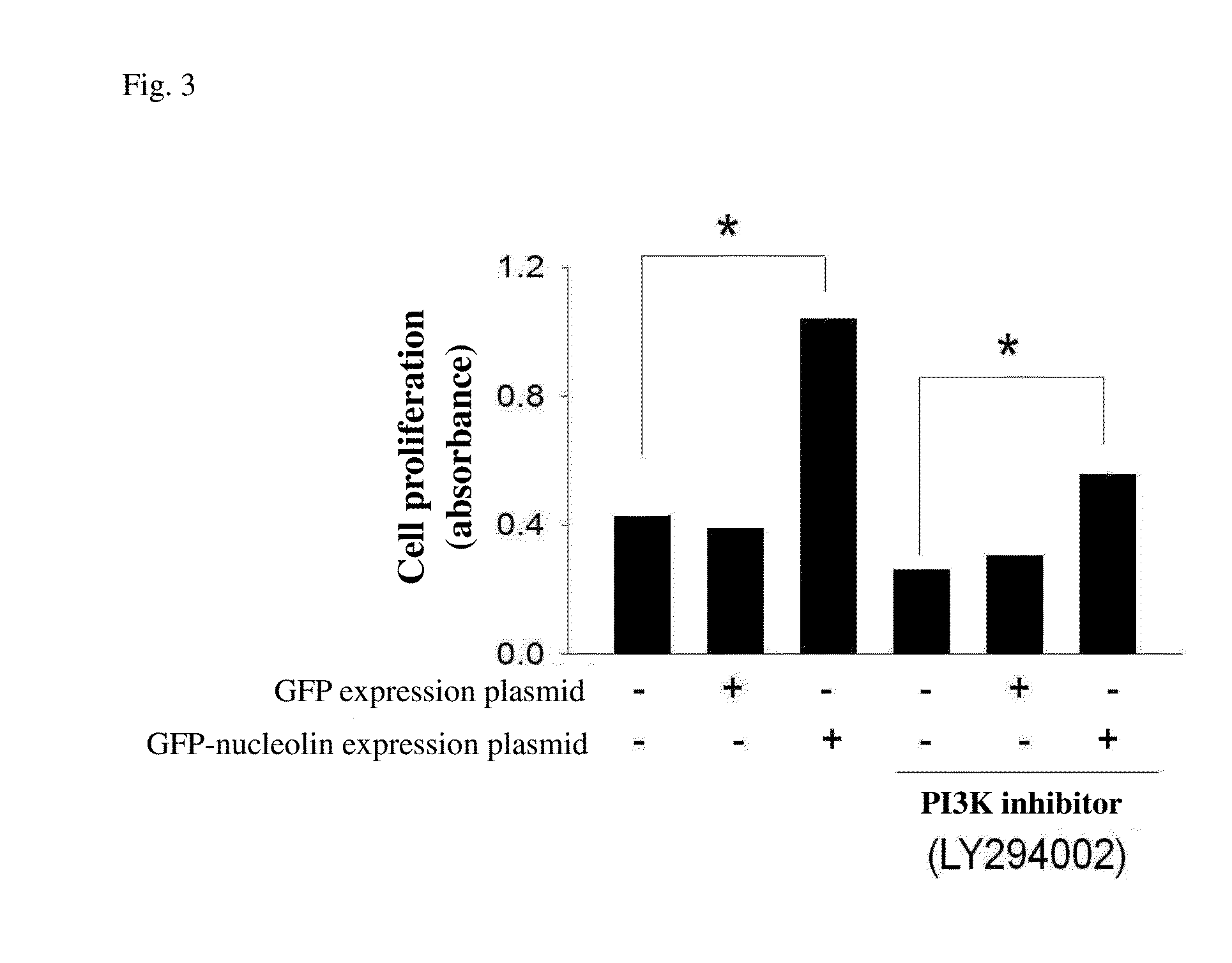
Effect of Nucleolin on Liver Cancer Cell Proliferation
[0061]cDNAs obtained from reverse transcription of RNA extracted from liver cancer cells (SK-Hep-1 cell line) were subjected to amplification of nucleolin by using forward primer (5′-atggtgaagctcgcgaaggcag-3′, SEQ ID NO: 2) and reverse primer (5′-ttcaaacttcgtcttctttccttg-3′, SEQ ID NO: 3) for nucleolin, and reverse transcription polymerase. The amplification product was inserted into green fluorescent protein (GFP) expression plasmid to form GFP-nucleolin expression plasmid. This GFP-nucleolin expression plasmid was transiently transfected into human liver cancer cells, so that the cells could express a lot of nucleolin protein. 6×10⁵ liver cancer cells were cultured in a 6-well sterile petri dish for 24 hours at 37° C. Then GFP-nucleolin expression plasmid and a control group of GFP expression plasmid were transfected into liver cancer cells by Opti-MEM transfection reagent (purchased from Gibco) in accordance with the reagent i...
PUM
 Abstract
Description
Claims
Application Information
 - R&D Engineer
- R&D Manager
- IP Professional
- Industry Leading Data Capabilities
- Powerful AI technology
- Patent DNA Extraction
Browse by: Latest US Patents, China's latest patents, Technical Efficacy Thesaurus, Application Domain, Technology Topic.
© 2024 PatSnap. All rights reserved.Legal|Privacy policy|Modern Slavery Act Transparency Statement|Sitemap