Systems and methods for enhanced implantation of electrode leads between tissue layers

a technology of electrode leads and tissue layers, applied in the field of systems and methods for implanting electrode leads for neuromuscular electrical stimulation, can solve the problems of spinal stabilization system dysfunction, back pain, and corrupt signal transduction, and achieve the effects of restoring muscle function to the lumbar spine, reducing the risk of a high stress location, and enhancing the implantation of electrode leads

Active Publication Date: 2018-01-11
MAINSTAY MEDICAL
View PDF3 Cites 104 Cited by
  • Summary
  • Abstract
  • Description
  • Claims
  • Application Information

AI Technical Summary

Benefits of technology

[0027]The present disclosure describes systems and methods for enhanced implantation of an electrode lead between tissue layers to reduce the risks of a high stress location on the lead or dislodgement. The lead may be configured to emit electrical energy from one or more electrodes disposed on the lead to stimulate target tissue to restore muscle function to the lumbar spine and treat, for example, low back pain. The systems and methods are expected to provide efficient implantation of the lead in a midline-to-lateral manner such that the implanted lead may be secured within the patient and used to restore muscle function of local segmental muscles associated with the lumbar spine stabilization system without disruption of the electrode lead post-implantation due to surrounding anatomical structures.

Problems solved by technology

It is believed that some cases of back pain are caused by abnormal mechanics of the spinal column.
Ligament injury, fatigue, and viscoelastic creep may corrupt signal transduction.
It is believed that in some patients with back pain, the spinal stabilization system is dysfunctional.
With soft tissue injury, mechanoreceptors may produce corrupted signals about vertebral position, motion, or loads, leading to an inappropriate muscle response.
In addition, muscles themselves may be injured, fatigued, atrophied, or lose their strength, thus aggravating dysfunction of the spinal stabilization system.
Conversely, muscles can disrupt the spinal stabilization system by going into spasm, contracting when they should remain inactive, or contracting out of sequence with other muscles.
As muscles participate in the feedback loop via mechanoreceptors in the form of muscle spindles and golgi tendon organs, muscle dysfunction may further compromise normal muscle activation patterns via the feedback loops.
Normally, load transmission is painless.
Over time, dysfunction of the spinal stabilization system is believed to lead to instability, resulting in overloading of structures when the spine moves beyond its neutral zone.
High loads can lead to inflammation, disc degeneration, facet joint degeneration, and muscle fatigue.
Since the endplates and annulus have a rich nerve supply, it is believed that abnormally high loads may be a cause of pain.
Load transmission to the facets also may change with degenerative disc disease, leading to facet arthritis and facet pain.
There are a number of problems associated with current implants that aim to restore spine stabilization.
First, it is difficult to achieve uniform load sharing during the entire range of motion if the location of the optimum instant axis of rotation is not close to that of the motion segment during the entire range of motion.
Second, cyclic loading of dynamic stabilization implants may cause fatigue failure of the implant, or the implant-bone junction, e.g. screw loosening.
Third, implantation of these systems requires surgery, which may cause new pain from adhesions, or neuroma formation.
Moreover, surgery typically involves cutting or stripping ligaments, capsules, muscles, and nerve loops, which may interfere with the spinal stabilization system.
In patients with spinal cord injury, the sensory nervous system is usually damaged as well as the motor system, and thus the afflicted person loses proprioception of what the muscle and limbs are doing.
Such a process can result in complications depending on the insertion site due to anatomical structures surrounding the target implantation site, impeding the insertion path.
There is a risk that electrode lead conductors may experience a tight bend near the location where the lead enters the thoracolumbar fascia when the lead is inserted within the body near the lateral edge of the spine.
Such a tight bend may lead to dislodgement of the electrode lead and / or fracture, thereby preventing proper therapy delivery.
The difference in directions of the superficial and deep fascia near the insertion site at the lateral edge of the spine may increase the risk of a high stress location on the lead.

Method used


Image

  • Systems and methods for enhanced implantation of electrode leads between tissue layers
  • Systems and methods for enhanced implantation of electrode leads between tissue layers
  • Systems and methods for enhanced implantation of electrode leads between tissue layers

Examples


Embodiment Construction

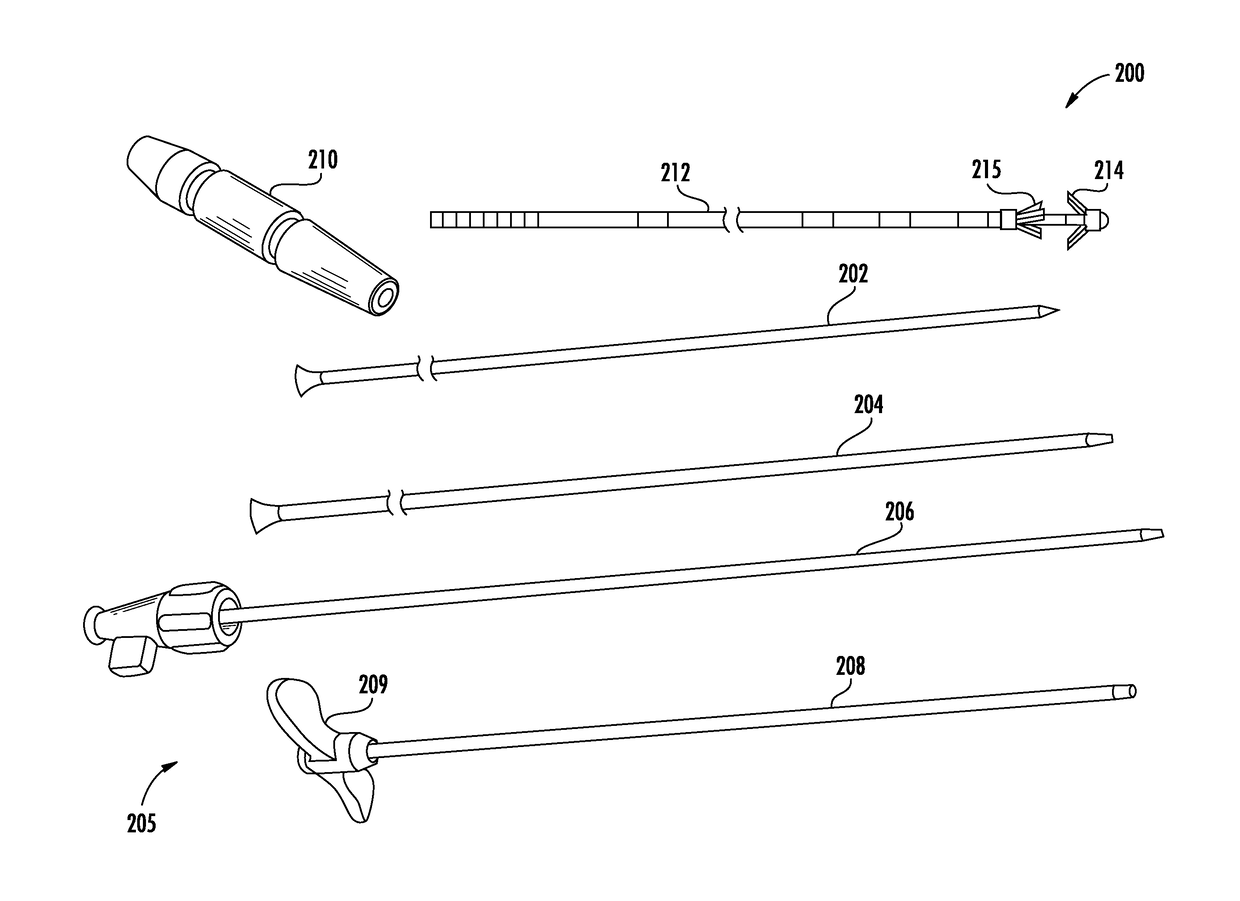
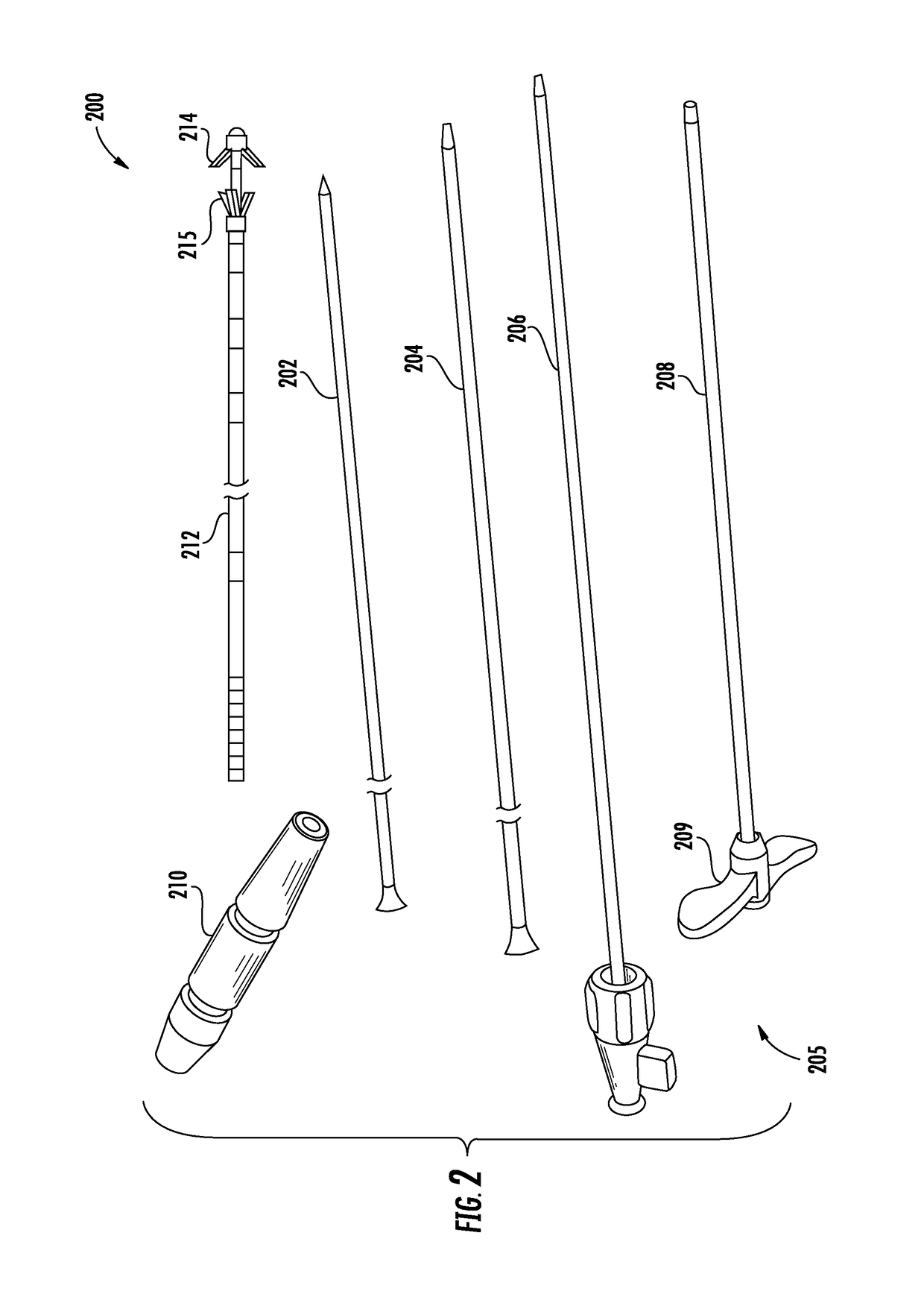
[0044]The systems and methods of the present disclosure may provide efficient implantation of an electrode lead in a midline-to-lateral manner such that the implanted lead may be secured within the patient and used to restore muscle function of local segmental muscles associated with the lumbar spine stabilization system without disruption of the electrode lead post-implantation due to surrounding anatomical structures. In accordance with the principles of the present disclosure, the systems and methods may be optimized for use in restoring muscle function to the lumbar spine to treat, for example, low back pain.

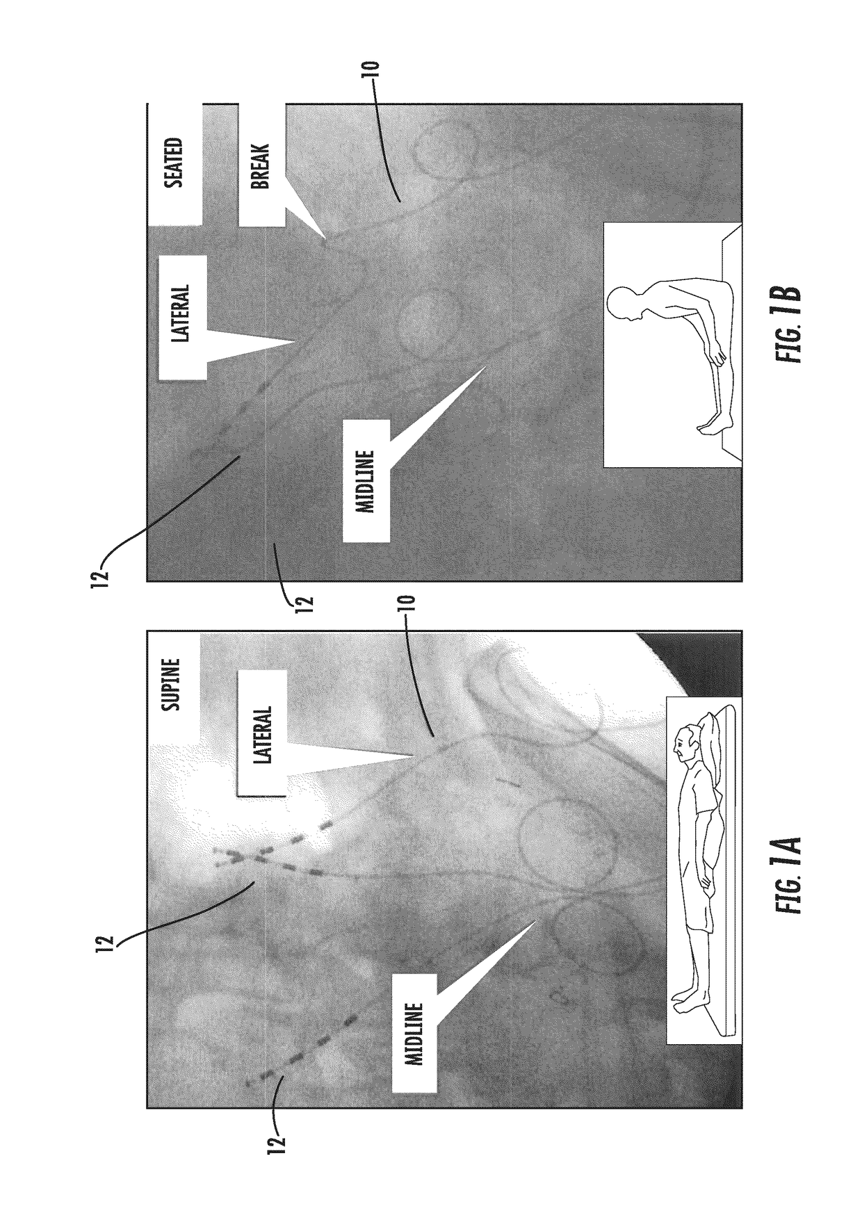
[0045]Referring to FIGS. 1A and 1B, a comparison of traditional implantation methods and the exemplary method in accordance with the principles of the present disclosure is provided. FIGS. 1A and 1B illustrate x-ray images of the lumbar region of a cadaver with electrode lead 10 and electrode leads 12 implanted therein. Electrode lead 10 was implanted via traditional methods...


PUM


Abstract

Systems and methods for enhanced implantation of an electrode lead for neuromuscular electrical stimulation of tissue associated with control of the lumbar spine for treatment of back pain, in a midline-to-lateral manner are provided. The implanted lead may be secured within the patient and used to restore muscle function of local segmental muscles associated with the lumbar spine stabilization system without disruption of the electrode lead post-implantation due to anatomical structures.

Description

I. FIELD OF THE INVENTION[0001]This application generally relates to systems and methods for implanting electrode leads for neuromuscular electrical stimulation, including stimulation of tissue associated with control of the lumbar spine for treatment of back pain.II. BACKGROUND OF THE INVENTION[0002]The human back is a complicated structure including bones, muscles, ligaments, tendons, nerves and other structures. The spinal column has interleaved vertebral bodies and intervertebral discs, and permits motion in several planes including flexion-extension, lateral bending, axial rotation, longitudinal axial distraction-compression, anterior-posterior sagittal translation, and left-right horizontal translation. The spine provides connection points for a complex collection of muscles that are subject to both voluntary and involuntary control.[0003]Back pain in the lower or lumbar region of the back is common. In many cases, the cause of back pain is unknown. It is believed that some ca...

Claims


Application Information

Patent Timeline
Patent Type & Authority Applications(United States)
IPC IPC(8): A61B17/34A61B18/00
CPCA61B17/3468A61B2018/00339A61B18/00A61N1/0551A61N1/0558
Inventor SHIROFF, JASON ALANDEMORETT, HENRYBECK, JOHNHAUSCHILD, SIDNEY
Owner MAINSTAY MEDICAL
Why Eureka
Try Eureka