
Apparatus for performing nanoparticle-assisted external beam radiotherapy and method carried out using said apparatus

a technology of external beam and apparatus, which is applied in the field of apparatus for performing nanoparticle-assisted external beam radiotherapy, can solve the problems of limiting the scope of optimizing the therapeutic protocol, affecting estimation, and inability to measure the dose delivered to the target region of the body

Active Publication Date: 2019-06-13
COMMISSARIAT A LENERGIE ATOMIQUE ET AUX ENERGIES ALTERNATIVES
View PDF0 Cites 0 Cited by
  • Summary
  • Abstract
  • Description
  • Claims
  • Application Information

AI Technical Summary

Benefits of technology

The invention aims to improve X-ray external beam radiotherapy using nanoparticles. It aims to allow for the measurement of the delivered X-ray dose and, preferably, the mass of the nanoparticles within the patient's body.

Problems solved by technology

The technical problem addressed in this patent text is the uncertainty in the estimation of the dose of ionizing radiation delivered to the target region of the body during external beam radiotherapy, which limits the scope for optimizing the therapeutic protocol. The text proposes the use of metal nanoparticles to enhance the therapeutic effect of ionizing radiation and improve the accuracy of dose estimation.

Method used


Image

  • Apparatus for performing nanoparticle-assisted external beam radiotherapy and method carried out using said apparatus
  • Apparatus for performing nanoparticle-assisted external beam radiotherapy and method carried out using said apparatus
  • Apparatus for performing nanoparticle-assisted external beam radiotherapy and method carried out using said apparatus

Examples


Embodiment Construction

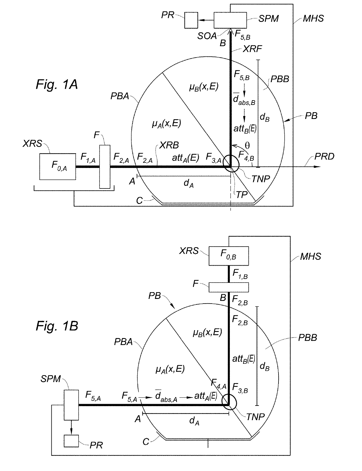
[0046]As illustrated on FIGS. 1A and 1B, an external radiotherapy apparatus according to the invention comprises:

[0047]an X-ray source XRS, such as a conventional radiotherapy X-ray tube (alternatively, an external X-ray source may be fitted to the apparatus), adapted for generating an X-ray beam XRB propagating along a propagation direction PRD. Typically, the X-ray source XRS operates in the orthovoltage (200-500 keV) or megavoltage (1-6 MeV) range.

[0048]An X-ray filter F, disposed on the propagation path of the X-ray beam—the design of this filter will be discussed extensively.

[0049]An X-ray spectrometer SPM, sensible in all or part of the 7-130 keV spectral range. Indeed, for all nanoparticle materials of interest, Kα and Kβ fluorescence lines are comprised in the 10-100 keV, and it is preferable that the spectrometer is sensitive in a region of at least ±30% of this range.

[0050]The spectrometer SPM has an optical axis SOA which forms an angle θ with the propagation direction PR...


PUM


Abstract

An apparatus for performing nanoparticle-assisted external beam radiotherapy includes an X-ray spectrometer having an optical axis, an X-ray filter, and a mobile holding structure suitable for holding the X-ray filter and the X-ray spectrometer in a first and a second relative position, and for switching from the first to the second relative position while simultaneously allowing the positioning of a patient body part in a target region including a target point; wherein: the first and second relative positions of the X-ray filter and the X-ray spectrometer are such that an X-ray beam emitted from the X-ray source crosses the X-ray filter before reaching the target point, a propagation direction of the X-ray beam forming an angle θ different from 0° and 180° with the optical axis of the X-ray spectrometer; and the second relative position is obtained by inverting the relative positions of the X-ray filter and of the X-ray spectrometer with respect to the patient body part. A method of determining an X-ray dose delivered at a region of a patient body using the apparatus is provided.

Description


Claims


Application Information

Patent Timeline
Owner COMMISSARIAT A LENERGIE ATOMIQUE ET AUX ENERGIES ALTERNATIVES
Learn More