

Detecting, localizing, and targeting internal sites in vivo using optical contrast agents
a technology of optical contrast and internal sites, applied in the direction of catheters, instruments, diagnostics using spectroscopy, etc., can solve the problems of inability to possess strong distinguishing features, and inability to detect the presence of key molecules in many disease processes. to achieve the effect of rapid imaging, localization, positioning and targeting of tissu
- Summary
- Abstract
- Description
- Claims
- Application Information
AI Technical Summary
Benefits of technology
Problems solved by technology
Method used
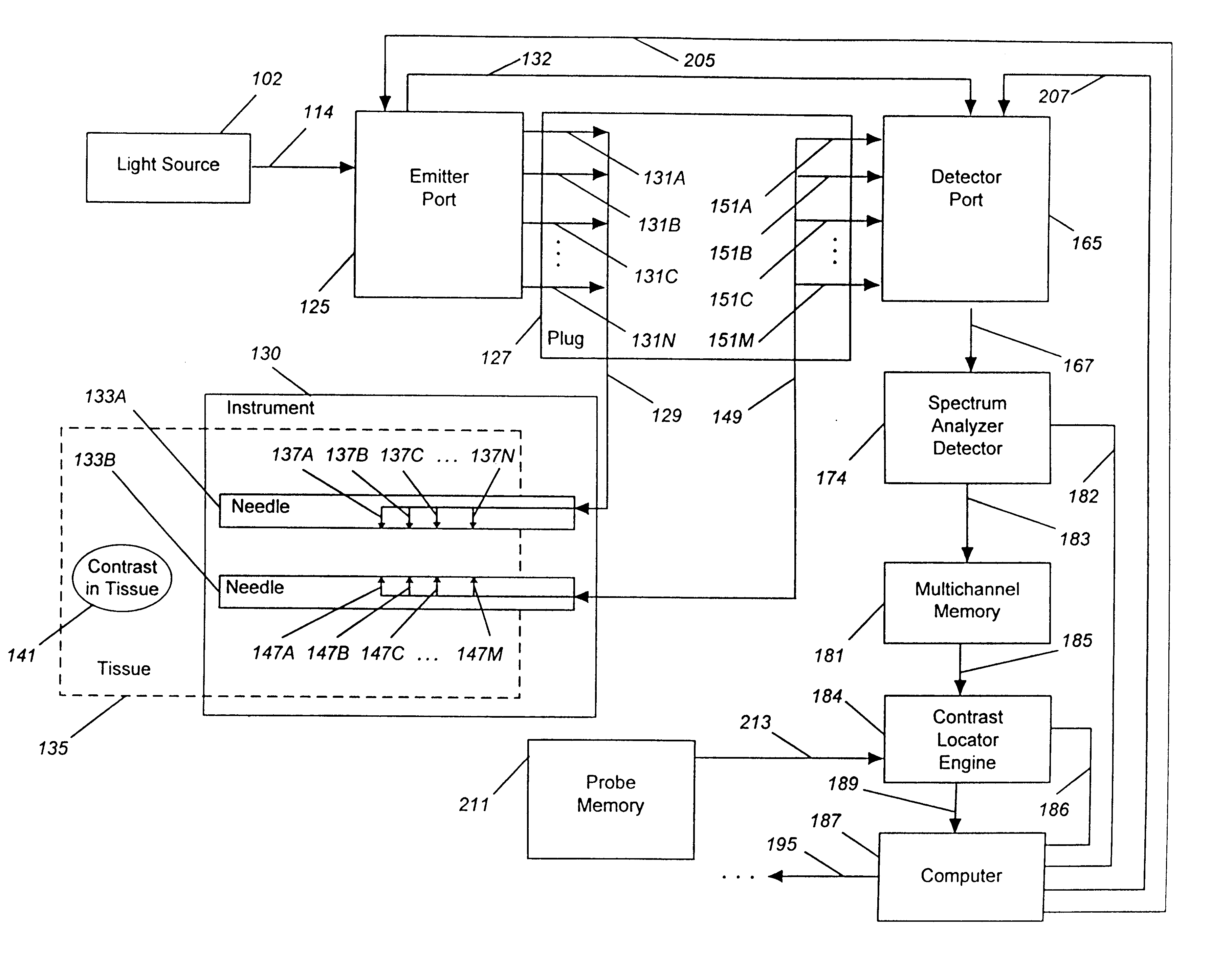
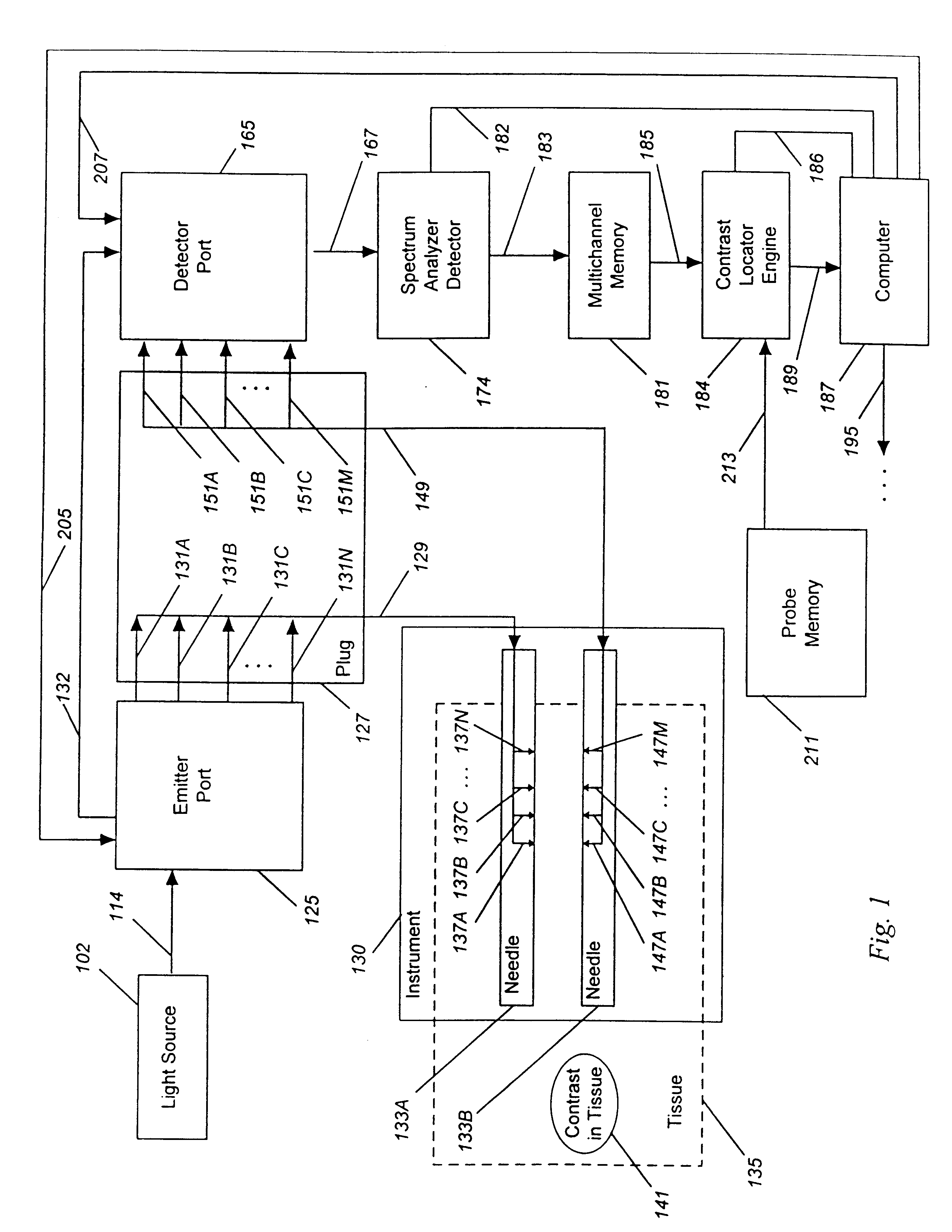
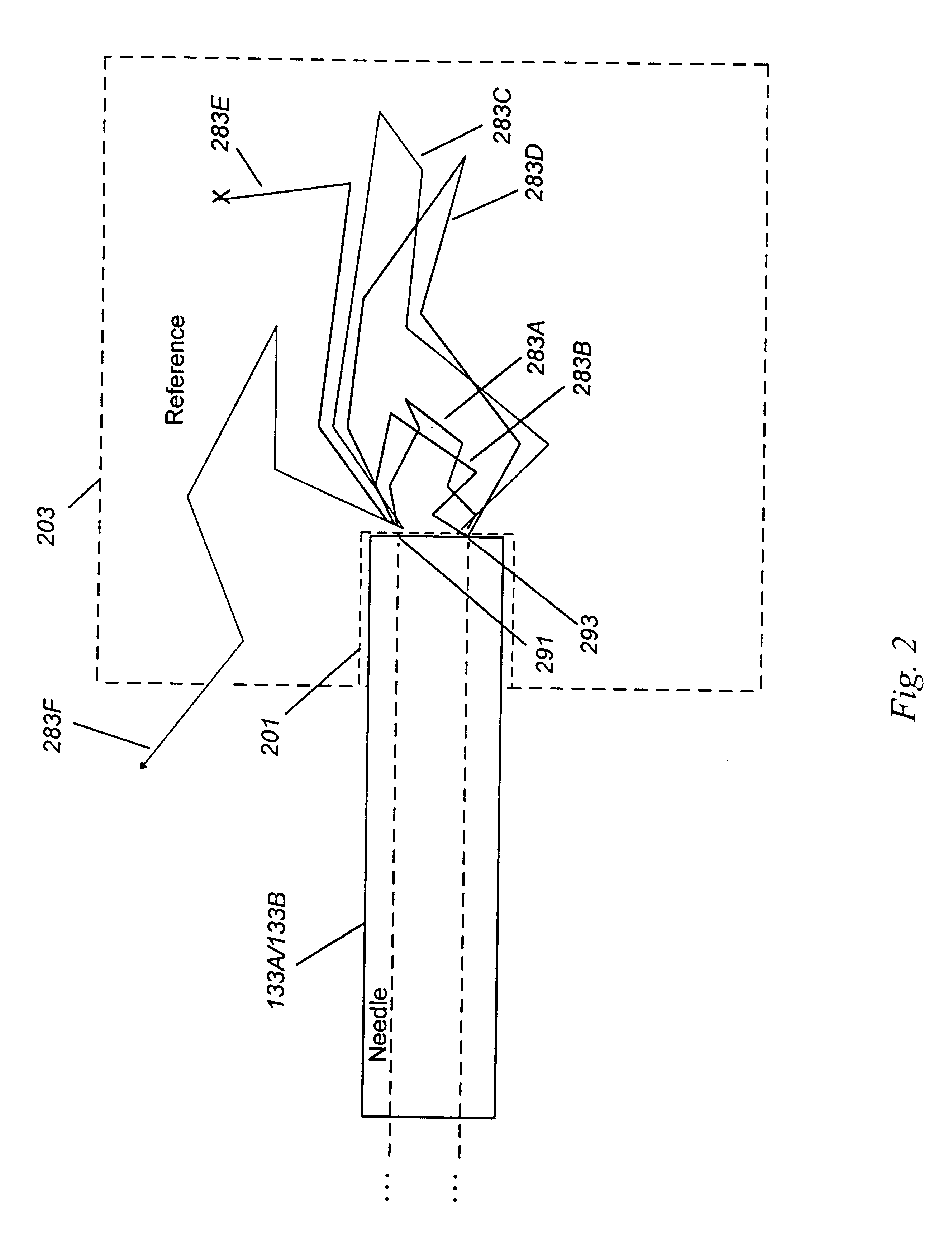
Image

Examples
example 1
Targeting of a Needle in a Human Tissue to a Contrast-Positive Site
Localization of a tissue at which biopsy should be collected may be assisted using a contrast that collects at the target site. Such a device would be useful for prostate cancer. One in five men will develop the disease, which exceeds even the incidence of breast cancer in women. The standard method of diagnosis, prostate biopsy, has a 20% chance of missing a cancer that is present. Thus, repeat biopsy tests are frequently required. Currently, less that 20% of all prostate tumors can be felt during examination, and as prostate cancer is not well seen by current imaging methods, prostate biopsies are in effect performed blindly.
Many methods of targeting a contrast agent to prostate cancer can be utilized. For example, as discussed earlier, it is known that certain cell types possess surface markers such that cancer cells may have surface receptors that their neighbor cells do not. This allows for contrast agents to be...
example 2
Surface Detection and Imaging of Isosulfan-Positive Areas Another example of a targeted procedure is biopsy of lymph nodes for breast cancer. Breast cancer is the most common cancer in women worldwide, second only to cervical cancer, and is a leading cause of death in women.
The treatment of breast cancer requires determination of the grade and spread of the tumor, a process called staging. Determination of spread to the lymph nodes is a key to proper treatment selection and increased survival. When breast cancer is diagnosed, the sentinel lymph node is identified and biopsied. Sentinel nodes are the main lymph node draining the tumor. They are currently identified by injection of a radioactive substance, which is traced using a Geiger counter (e.g., U.S. Pat. No. 5,846,513 discussed under Background) with or without the injection of blue contrast. Sentinel breast nodes can be marked by injection of the tumor with blue contrast, as the contrast migrates to the sentinel lymph node or ...
example 3
Multicolor Contrast Labeling
Dual or multiple contrast labeling is a technique frequently used in cytology and other ex vivo laboratory disciplines. In this technique, different contrast agents are added to the same sample, in order to extract additional information. This approach can also be applied in vivo using the present method.
For example, many tissues may take up a first contrast agent, as these tissues have a receptor for the first contrast agent. In addition, a different combination of tissues may take up a second contrast agent, as these different tissues have a receptor for the second marker. A cancer may be identified as having both receptors, or the presence of one without the presence of the second. In either case, the ability to detect, localize, image, and target using two or more contrast agents simultaneously may be important.
As a test of this, the probe was used to differentiate regions containing no exogenous contrast agent from regions containing the a red contra...
PUM
 Abstract
Description
Claims
Application Information