Liposome containing hydrophobic iodine compound and X-ray contrast medium for radiograph comprising the liposome

a technology of hydrophobic iodine and liposome, which is applied in the field of liposome, can solve the problems of low resolution of arteriosclerosis, inability to detect arteriosclerosis, and inability to meet the requirements of a satisfactory method for diagnosing the progress of arteriosclerosis

Inactive Publication Date: 2006-03-07
FUJIFILM HLDG CORP +1
View PDF5 Cites 8 Cited by
  • Summary
  • Abstract
  • Description
  • Claims
  • Application Information

AI Technical Summary

Benefits of technology

[0011]A further object of the present invention is to provide an X-ray contrast medium which enables sharp imaging of a biological environment of a tissue or a lesion in which macrophages localize such as in liver, spleen, air vesicle, lymph node, lymph vessel, renal epithelium or the like. A still further object of the present invention is to provide a contrast medium which enables identification of a site in which tumor, inflammation, or infection is progressing.

Problems solved by technology

However, no satisfactory method is available for diagnosing progress of arteriosclerosis at an early stage before the onsets of the aforementioned diseases.
However, by these methods, it is almost impossible to detect arteriosclerosis at an early stage, especially constriction of coronary artery, that causes myocardial infarction or angina pectoris, at an early stage before the onset of these diseases.
However, these methods have been mainly developed for detection of tumors, and accordingly, they have a problem of a low resolution of arteriosclerotic lesions.
In addition, the methods require expensive and large-scale apparatuses, which limits employable hospitals and general applicability.
However, for employment of these methods, it is necessary to arterially insert an ultrasonic oscillator or an endoscope attached to an end of a catheter, which may result in serious physical stress and heaviness as well as a risk of a patient.
Therefore, although these methods have been used therapeutically for patients after the attack of myocardial infarction and the like or as secondary prophylaxis, they cannot be used for a diagnostic purpose to know as to presence or absence or a degree of progress of arteriosclerosis in a patient before onset.
However, these compounds are rapidly excreted from such lumens without interaction with a tissue or a lesion, and therefore, they are not useful for diagnosing a tissue or a lesion in more detail.
Moreover, these methods can only detect a lesion where constriction progresses 50% or more and fail to detect a lesion before the onset of attack of an ischemic disease.
However, these methods for formulation are not sufficient in efficiency and selectivity for a purpose of selective radiography of a tissue or a lesion.
However, the methods using these formulations are not satisfactory in efficiency and selectivity for a purpose of selective contrast of vascular diseases, and no example thereof is reported in which vascular diseases are imaged by utilizing X-ray irradiation.

Method used


Image

  • Liposome containing hydrophobic iodine compound and X-ray contrast medium for radiograph comprising the liposome
  • Liposome containing hydrophobic iodine compound and X-ray contrast medium for radiograph comprising the liposome
  • Liposome containing hydrophobic iodine compound and X-ray contrast medium for radiograph comprising the liposome

Examples


example 1

Preparation of Culture System of Vascular Smooth Muscle Cells of which Proliferation is Activated by Foam Macrophages

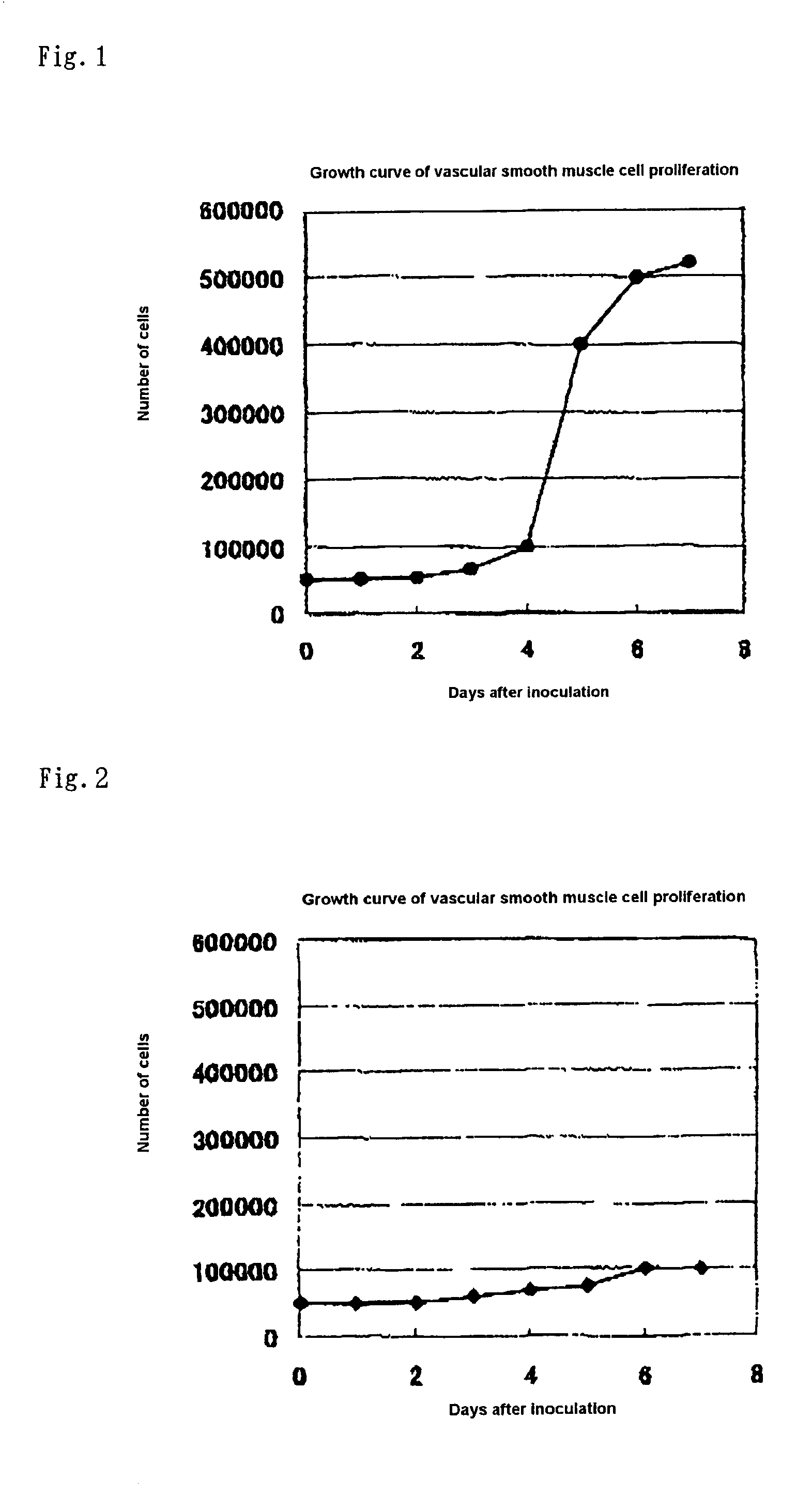
[0056]Vascular smooth muscle cells were isolated from mouse aorta endothelium (“Tissue Culture Method”, 10th Edition, ed. by the Japanese Tissue Culture Association, published by Kodansha, 1998). The isolated vascular smooth muscle cells were suspended in 10% FBS Eagle's MEM (GIBCO, No. 11095-080) and inoculated in wells of a 12-well microplate (FALCON, No. 3503). The number of the cells in each well was adjusted to 10,000 cells. The cells were cultured for 3 days under conditions of 37° C. and 5% CO2.

[0057]Then, foamed mouse peritoneal macrophages were prepared according to the method described in Biochimica Biophysica Acta, 1213, 127 (1994). 200,000 cells of the foam macrophages were separated and inoculated on an insert cell (FALCON, No. 3180) placed on each well of the microplate where the vascular smooth muscle cells were cultured on the bottom surface. The cells...

example 2

Verification of Expression of Scavenger Receptors on Vascular Smooth Muscle Cells

[0058]It is known that vascular smooth muscle cells in an arteriosclerotic lesion express scavenger receptors on their surfaces to take up oxidized LDL (Biochem. Phamacol., 15:57 (4), 383 (1999); Exp. Mol. Pathol., 64 (3), 127–45, 1997). The vascular smooth muscle cells of the culture system of FIG. 1 were immunostained by using mouse scavenger receptor antibodies. As a result, although the expression was not observed on the vascular smooth muscle cells on the 3rd day from the inoculation, clear staining was observed on the 6th day from the inoculation. When the foam macrophages on the cell filter was also similarly immunostained, clear staining was also observed.

example 3

Uptake of Oxidized LDL by Vascular Smooth Muscle Cells

[0059]In the culture system of FIG. 1, 125I-labeled oxidized LDL was added to the medium for the vascular smooth muscle cells on the 3rd day and 6th day from the inoculation. 125I taken up into the cells was counted 24 hours after each addition. The results are shown in Table 1. Clear difference in uptake amount was observed between the results on the 3rd day and 6th day.

[0060]

TABLE 1Uptake of 125I-oxLDLDays(× 1,000 cpm)3 Days after inoculation0.52 ± 0.116 Days after inoculation2.2 ± 0.4

[0061]The above results indicate that the vascular smooth muscle cells cultured in the aforementioned cell culture system had properties similar to those of smooth muscle cells in a lesion of arteriosclerosis, restenosis or the like. Because the vascular smooth muscle cells cultured in the culture system in FIG. 1 had properties of healthy blood vessel until the 3rd day from the inoculation and properties of smooth muscle cells in an arteriosclero...


PUM

PropertyMeasurementUnit
thicknessaaaaaaaaaa
sizeaaaaaaaaaa
sizeaaaaaaaaaa

Abstract

A liposome containing a hydrophobic iodine compound represented by the following general formula (I) as a membrane component:R1—CO2—R2wherein R1 represents a substituted or unsubstituted 2,3,5-triiodophenyl group or a substituted or unsubstituted 3,4,5-triiodophenyl group; and R2 represents a hydrocarbon group having 10 or more carbon atoms, and an X-ray contrast medium, which comprises said liposome which is used for radiography of a vascular disease.

Description

FIELD OF THE INVENTION[0001]The present invention relates to a liposome. More specifically, the invention relates to a liposome that contains a particular class of hydrophobic iodine compound and can be utilized for a method for imaging a lesion in contrast to non-pathological site by selectively accumulating the hydrophobic iodine compound in the lesion. The present invention also relates to an X-ray contrast medium which selectively accumulates in a tissue or lesion, in which macrophages localize, and enables imaging thereof in contrast to the other sites.RELATED ART[0002]In the modern society, especially in the societies of advanced countries, opportunities of ingesting high calorie and high fat diet are increasing. For this reason, mortalities due to ischemic diseases resulting from arteriosclerosis (heart diseases such as myocardial infarction and angina pectoris, cerebrovascular diseases such as cerebral infarction and cerebral hemorrhage) have been increasing. Therefore, it h...

Claims


Application Information

Patent Timeline
Patent Type & Authority Patents(United States)
IPC IPC(8): A61K49/00A61K9/127A61K49/04
CPCA61K49/0466
Inventor KITAGUCHI, HIROSHIAIKAWA, KAZUHIRO
Owner FUJIFILM HLDG CORP
Why Eureka
Try Eureka