Surgical perforation device with electrocardiogram (ECG) monitoring ability and method of using ECG to position a surgical perforation device

a perforation device and surgical technology, applied in the field of surgical perforation devices with ecg monitoring ability and method of using ecg to position the surgical device, can solve the problems of rapid increase in intracellular temperature, subsequent cell lysis, and vaporization of intracellular water

Active Publication Date: 2007-09-18
BOSTON SCI MEDICAL DEVICE LTD
View PDF37 Cites 152 Cited by
  • Summary
  • Abstract
  • Description
  • Claims
  • Application Information

AI Technical Summary

Problems solved by technology

Further, when the material to be cut comprises cellular tissue, the energy delivery device is operable to deliver sufficient energy to the tissue to result in a rapid increase in the intracellular temperature causing vaporization of intracellular water and subsequent cell lysis.

Method used


Image

  • Surgical perforation device with electrocardiogram (ECG) monitoring ability and method of using ECG to position a surgical perforation device
  • Surgical perforation device with electrocardiogram (ECG) monitoring ability and method of using ECG to position a surgical perforation device
  • Surgical perforation device with electrocardiogram (ECG) monitoring ability and method of using ECG to position a surgical perforation device

Examples


Embodiment Construction

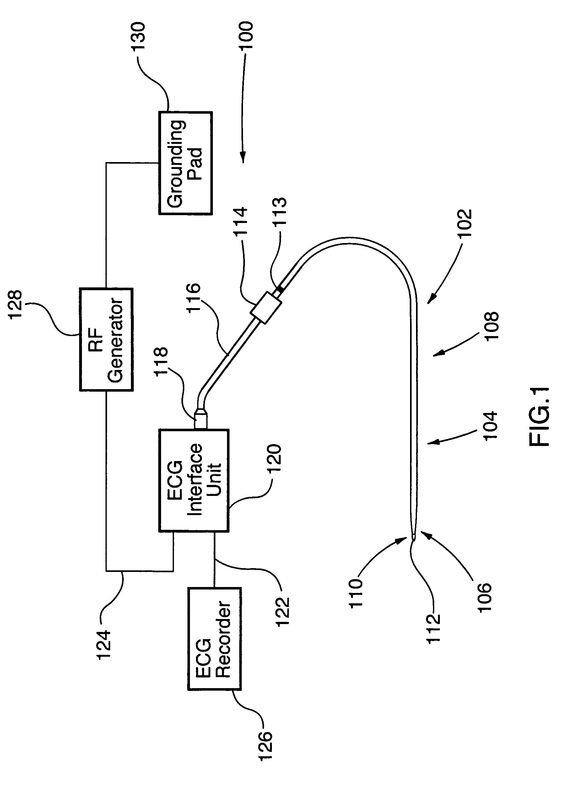
[0035]FIG. 1 illustrates an embodiment of an electrosurgical perforation device 102 in accordance with the invention in an electrosurgical system 100. Device 102 comprises an elongate member 104 having a distal region 106, and a proximal region 108. Distal region 106 is adapted to be inserted within and along a lumen of a body of a patient, such as a patient's vasculature, and maneuverable therethrough to a desired location proximate to material such as tissue to be cut.

[0036]The elongate member 104 is typically tubular in configuration, having at least one lumen 200 extending from proximal region 108 to distal region 106. Elongate member 104 is preferably constructed of a biocompatible polymer material that provides column strength to device 102. The elongate member 104 is sufficiently stiff to permit a dilator 710 and a soft guiding sheath 700 to be easily advanced over device 102 and through a perforation. Examples of suitable materials for the tubular portion of elongate member ...


PUM


Abstract

A device with a functional tip containing at least one active electrode capable of creating a controlled perforation in body tissue through the application of energy (e.g. Radio Frequency (RF)) is described. The position of the tip of the device can be determined in response to ECG measured at the tip and determined by a monitor coupled to the device. The device is useful to remove or perforate unwanted tissue in a controlled manner in any location in the body, particularly in the atrial septum for controlled transseptal puncture. In this application, the device is introduced into the right atrium, and the functional tip is then positioned against the atrial septum. ECG is used to locate the region of the fossa ovalis on the atrial septum. Energy is applied to create the perforation and ECG is monitored to determine if the perforation was created in a desired location.

Description

TECHNICAL FIELD[0001]The invention relates to a surgical perforation device with ECG monitoring ability and method of using ECG to position the surgical device. More specifically, the invention relates to a device and method for creating a controlled perforation in the atrial septum while using ECG to locate the fossa ovalis region on the atrial septum, and delivering a dilator and guiding sheath to the left atrium through the perforation over the surgical device.BACKGROUND OF THE ART[0002]Electrosurgical devices perforate or cut tissues when radio frequency (RF) electrical energy rapidly increases tissue temperature to the extent that the intracellular fluid becomes converted to steam, inducing cell lysis as a result of elevated pressure within the cell. The radio frequency range lies between 10 kHz and 300 MHz, but electrosurgical devices usually operate at a frequency between 400 kHz and 550 kHz. This technology can be used to create perforations in different types of tissue, suc...

Claims


Application Information

Patent Timeline
Patent Type & Authority Patents(United States)
IPC IPC(8): A61B18/18A61B18/14A61B19/00
CPCA61B18/1492A61B2018/00351A61B2018/00601A61B2018/00702A61B2018/00839A61B2019/464A61B2218/002A61B2090/064
Inventor VISRAM, NAHEEDSHAH, KRISHAN
Owner BOSTON SCI MEDICAL DEVICE LTD
Why Eureka
Try Eureka