Eureka AIR delivers breakthrough ideas for toughest innovation challenges, trusted by R&D personnel around the world.

Meningeal-derived stem cells

a technology of stem cells and meningeal cells, applied in the field of stem cells, can solve the problems of ethical and political considerations, inability to obtain purified populations of desired cell types, and inability to use these cells, etc., and achieves the effects of high degree of developmental plasticity, robust self-renewal, and easy propagation in cultur

Inactive Publication Date: 2008-08-12
OGLE ROY C +1
View PDF5 Cites 3 Cited by
  • Summary
  • Abstract
  • Description
  • Claims
  • Application Information

AI Technical Summary

Benefits of technology

[0007]One aspect of the present invention is directed to stem cells derived from the dura mater, the pia mater or the arachnoid mater, as well as methodologies for isolation, differentiation and explantation of these cells. Meningeal tissue may be explanted (i.e., cells migrate out of adherent pieces of tissue) or enzymatically dissociated to yield primitive mesenchymal cells. The tissues may include those removed by biopsy from a patient or tissues removed aseptically from a fetus. These cells exhibit characteristics of “adult” stem cells or progenitors: robust self-renewal and a high degree of developmental plasticity. The stem cells may be readily propagated in culture (showing little senescence after 20 passages), and are capable of differentiating into various cell types. Thus, the meningeal-derived stem cells of the present invention are multipotent.
[0008]The meningeal stem cells of the present invention can be taken from a small biopsy, and rapidly expanded to large populations of cells using a specially defined media that maintains their undifferentiated state. Transformation to neural cells can be accomplished rapidly (i.e., within several hours) and bone and cartilage within two weeks, by adding factors that support and maintain these cell phenotypes. Schwann cells, adipocytes and fibroblasts may be rapidly produced, as well. In addition, the number of cells that transform to a neural morphology is between 90% and 95%. Thus, these cells may have particular utility in treating central nervous system (CNS) degenerative disorders and spinal cord injuries. The rapid proliferative capacity and high rate of neuronal differentiation makes these applications well-suited for the stem cells, although numerous other applications exist, as well.

Problems solved by technology

However, existing methods in the field have a number of major limitations that relate to obtaining purified populations of the desired cell types from pluripotent stem cells.
By way of example, embryonic stem cells pose interesting possibilities as several studies show that these cells are pluripotent, however, the use of these cells is mired in ethical and political considerations.
It is therefore likely that this technology will not be available for use in the near future.

Method used


Image

  • Meningeal-derived stem cells
  • Meningeal-derived stem cells
  • Meningeal-derived stem cells

Examples


example 1

Preparation of Nerve Cells

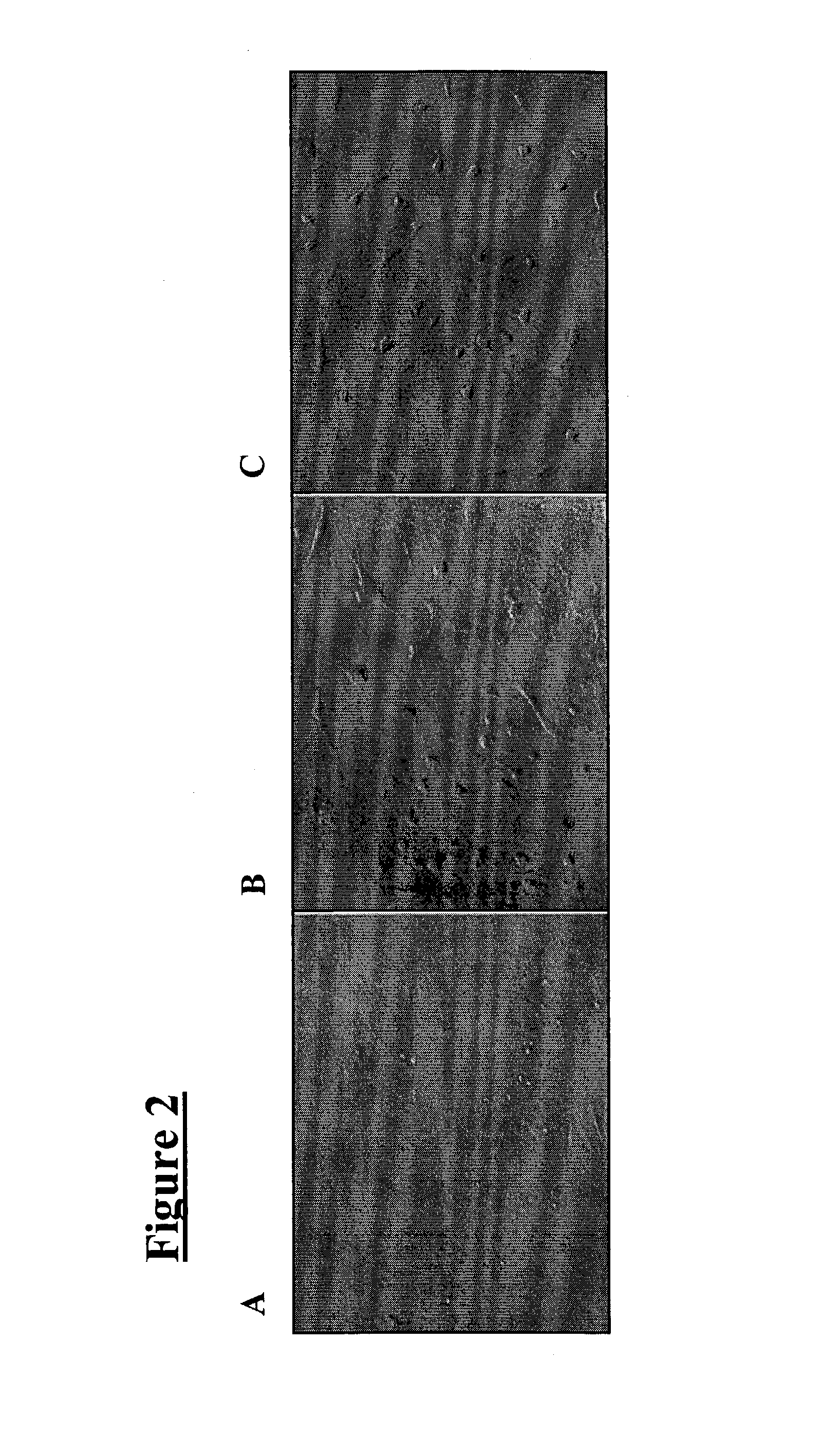
[0037]Cells from the meninges that are allowed to become 70% confluent (FIG. 2A) are susceptible to differentiating into nerve under two conditions: antioxidant treatment and steroid hormone treatment. When cells are exposed to a neuronal pre-induction media containing an antioxidant (DMEM, 20% FBS, 1 mM β-mercaptoethanol) for 24 hours, followed by treatment with neuronal induction media also containing an antioxidant (DMEM, 5 mM β-mercaptoethanol), the cells differentiate into neural-like cells within six hours (FIG. 2C). This can also be achieved with other antioxidants (i.e., reducing agents), such as butylated hydroxyanisole (BHA) (approximately 200 μM), dithiothreitol (DTT; i.e., Cleland's reagent), as well as dithioerythritol, tributylphosphine, iodoacetamide, tris-phosphine HCl, deoxythymidine-triphosphate trilithium salt, diethylthiatricarbocyanine perchlorate, diethylthiatricarbocyanine iodide and DECROLINE D (available from BASF Corporation; Mount...

example 2

Preparation of Bone Cells

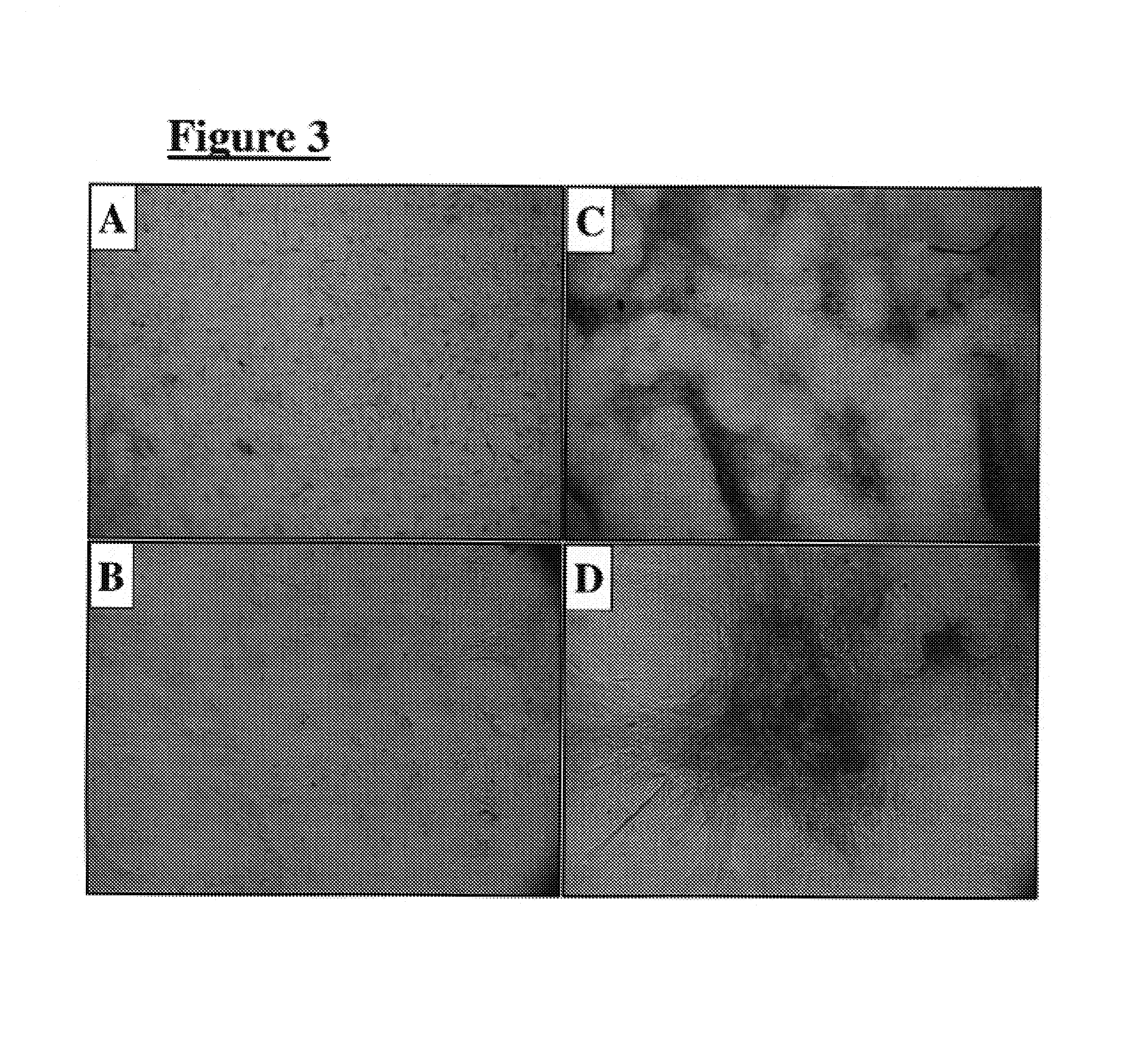
[0041]Dural cells were forced to adopt a bony phenotype by two separate methods. The first included plating these cells on a MATRIGEL or laminin substrate (100 μg / cm2). Plated cells expressed alkaline phosphatase (a differentiated bone marker) within seven days, and adopted an osteocytic morphology (FIGS. 3C & 3D). Untreated cells showed no staining for alkaline phosphatase (FIGS. 3A & 3B).

[0042]The second method involved exposing the cells to organic and inorganic phosphates. Inorganic phosphates included varying levels (i.e., 3-6 mM) of sodium phosphate and potassium phosphate, and organic phosphates included 10 mM β-glycerol phosphate. In addition, cells were exposed to 50 μg / ml ascorbic acid. Cells under these conditions also expressed alkaline phosphatase (data not shown).

example 3

Preparation of Cartilage

[0043]Cartilage development is fundamentally different from other tissues in that complex three-dimensional interactions are required to form nodules of cartilage in vitro. To accomplish this, a 10 μL volume of a 1×10⁷ cells / mL suspension was plated and allowed to attach to a tissue culture surface. This micromass culture differentiated into cartilage within two weeks when placed in media that contained 1X insulin-selenium-transferrin (ITS diluted 100-fold; available under the tradename ITS+ PREMIX from BD Biosciences) and 10 ng / ml transforming growth factor (TGF)-β1. The production of sulfated proteoglycan as demonstrated by Alcian Blue (available from Sigma-Aldrich Co.; St. Louis, Mo.) staining is indicative of chondrocytic differentiation (FIG. 4).


PUM

PropertyMeasurementUnit
volumeaaaaaaaaaa
concentrationaaaaaaaaaa
degree of developmental plasticityaaaaaaaaaa

Abstract

Stem cell lines derived from meningeal tissue such as dura mater, pia mater, or arachnoid mater, are disclosed. In various embodiments, the stem cell lines include greater than 80%, greater than 90%, or greater than 99% of pluripotent meningeal-derived stem cells. In other embodiments, the stem cell lines are induced to form nerve cells, bone cells, cartilage cells, Schwann cells, adipocytes, fibroblasts, or melanocytes with various inducing agents, such as antioxidants, steroid hormones, laminin, or various growth factors. Derivation of the current stem cell lines is accomplished through explantation or enzymatic dissociation of meningeal tissue, followed by expansion to large populations using growth media.

Description

RELATED APPLICATIONS[0001]This application claims the benefit of priority under 35 U.S.C. § 119 of provisional U.S. application Ser. No. 60 / 387,793, filed Jun. 11, 2002, the contents of which are hereby incorporated by reference.GOVERNMENT RIGHTS[0002]This invention was made with United States Government support under Grant No. SR01 DE010369 awarded by the National Institutes of Health. The United States Government has certain rights in the invention.FIELD OF THE INVENTION[0003]The present invention is directed to stem cells and methods of preparing populations of progenitor cells that differentiate into a preselected cell type with high efficiency.BACKGROUND OF THE INVENTION[0004]The brain and spinal cord are enclosed and protected by the meninges; tough and fibrous tissues comprising the dura mater and pia mater. Developmentally, these tissues form partly from the neural crest; a class of highly migratory and plastic cells that also form several diverse tissue types, such as bone,...

Claims


Application Information

Patent Timeline
Patent Type & Authority Patents(United States)
IPC IPC(8): C12N5/00C12N5/08C12N5/074C12N5/077C12N5/079C12N5/0793C12N5/0797
CPCC12N5/0607C12N5/0668C12N2533/90C12N2500/25C12N2500/38C12N2500/44C12N2500/46C12N2500/99C12N2501/01C12N2501/11C12N2501/115C12N2501/135C12N2501/15C12N2501/33C12N2501/385C12N2501/39C12N2501/70C12N2501/998C12N2533/50C12N2533/52C12N2533/54C12N2500/90
Inventor OGLE, ROY C.THOLPADY, SUNIL
Owner OGLE ROY C
Why Eureka
Eureka Blog
Learn More