Meningeal-derived stem cells
- Summary
- Abstract
- Description
- Claims
- Application Information
AI Technical Summary
Benefits of technology
Problems solved by technology
Method used
Image
Examples
example 1
Preparation of Nerve Cells
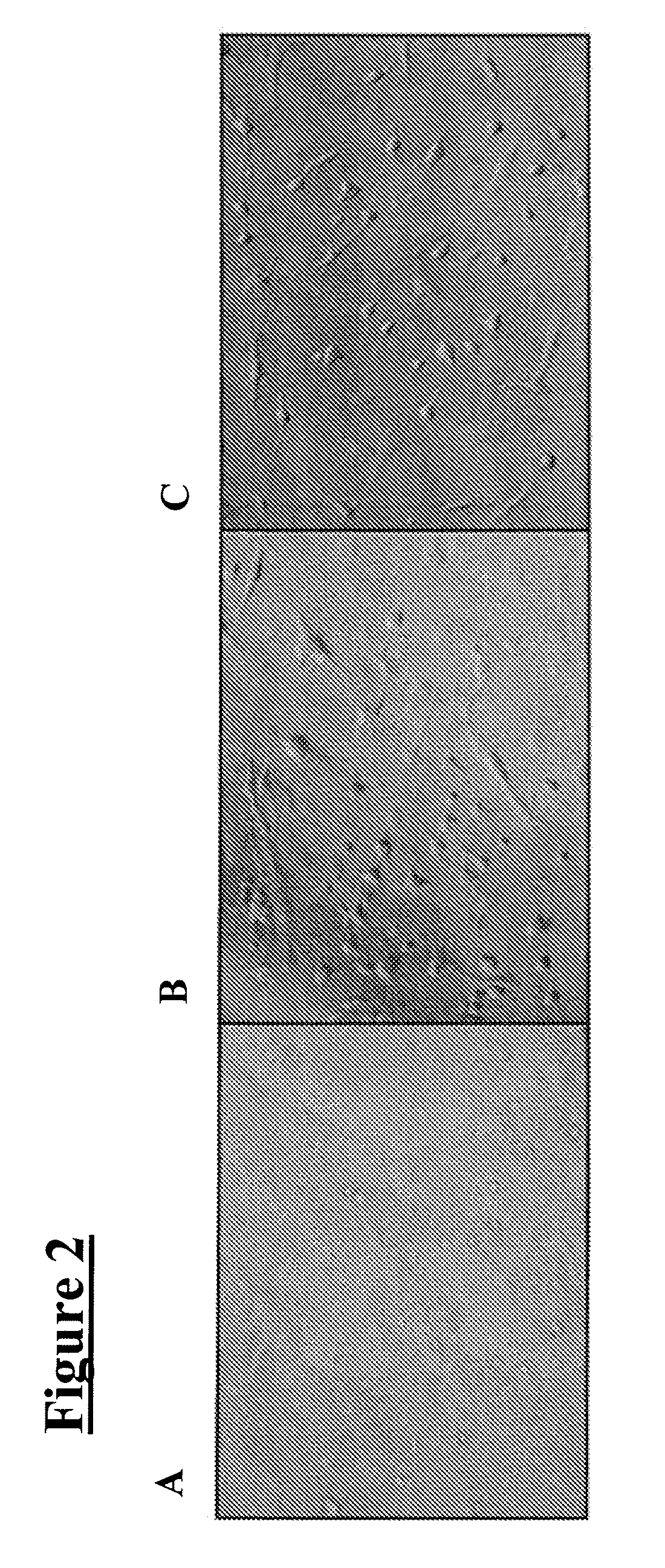
[0037] Cells from the meninges that are allowed to become 70% confluent (FIG. 2A) are susceptible to differentiating into nerve under two conditions: antioxidant treatment and steroid hormone treatment. When cells are exposed to a neuronal pre-induction media containing an antioxidant (DMEM, 20% FBS, 1 mM β-mercaptoethanol) for 24 hours, followed by treatment with neuronal induction media also containing an antioxidant (DMEM, 5 mM β-mercaptoethanol) the cells differentiate into neural-like cells within six hours (FIG. 2C). This can also be achieved with other antioxidants (i.e., reducing agents), such as butylated hydroxyanisole (BHA) (approximately 200 μM), dithiothreitol (DTT; i.e., Cleland's reagent), as well as dithioerythritol, tributylphosphine, iodoacetamide, tris-phosphine HCl, deoxythymidine-triphosphate trilithium salt, diethylthiatricarbocyanine perchlorate, diethylthiatricarbocyanine iodide and DECROLINE D (available from BASF Corporation; Moun...
example 2
Preparation of Bone Cells
[0041] Dural cells were forced to adopt a bony phenotype by two separate methods. The first included plating these cells on a MATRIGEL or laminin substrate (100 μg / cm2). Plated cells expressed alkaline phosphatase (a differentiated bone marker) within seven days, and adopted an osteocytic morphology (FIGS. 3C & 3D). Untreated cells showed no staining for alkaline phosphatase (FIGS. 3A & 3B).
[0042] The second method involved exposing the cells to organic and inorganic phosphates. Inorganic phosphates included varying levels (i.e., 3-6 mM) of sodium phosphate and potassium phosphate, and organic phosphates included 10 mM β-glycerol phosphate. In addition, cells were exposed to 50 μg / ml ascorbic acid. Cells under these conditions also expressed alkaline phosphatase (data not shown).
example 3
Preparation of Cartilage
[0043] Cartilage development is fundamentally different from other tissues in that complex three-dimensional interactions are required to form nodules of cartilage in vitro. To accomplish this, a 10 μL volume of a 1×10⁷ cells / mL suspension was plated and allowed to attach to a tissue culture surface. This micromass culture differentiated into cartilage within two weeks when placed in media that contained 1×insulin-selenium-transferrin (ITS diluted 100-fold; available under the tradename ITS+ PREMIX from BD Biosciences) and 10 ng / ml transforming growth factor (TGF)-β1. The production of sulfated proteoglycan as demonstrated by Alcian Blue (available from Sigma-Aldrich Co.; St. Louis, Mo.) staining is indicative of chondrocytic differentiation (FIG. 4).
PUM
 Abstract
Description
Claims
Application Information
 - R&D
- Intellectual Property
- Life Sciences
- Materials
- Tech Scout
- Unparalleled Data Quality
- Higher Quality Content
- 60% Fewer Hallucinations
Browse by: Latest US Patents, China's latest patents, Technical Efficacy Thesaurus, Application Domain, Technology Topic, Popular Technical Reports.
© 2025 PatSnap. All rights reserved.Legal|Privacy policy|Modern Slavery Act Transparency Statement|Sitemap|About US| Contact US: help@patsnap.com


