Detection of Anaplasma platys
a technology of anaplasma and platys, applied in the field of detection of anaplasma platys, can solve the problems of causing significant morbidity, limited specificity of current diagnostic tests that attempt to distinguish aph and apl, and the inability to detect anaplasma platys in time,
- Summary
- Abstract
- Description
- Claims
- Application Information
AI Technical Summary
Problems solved by technology
Method used
Image
Examples
example 1
Cloning of a p44 Homolog from Apl
[0079]A homolog of an Aph p44 gene from Apl was cloned from the blood of an infected dog. The blood sample was obtained from a dog residing on the Hopi Reservation in Arizona. Genomic DNA was isolated from 200 ul of whole blood using standard techniques (QiaAmp DNA Blood Miniprep Kit—Part #51104). Degenerate primers were designed to target the conserved regions of Aph p44, Anaplasma marginale msp2, Anaplasma ovis msp2, and Anaplasma centrale msp2 genes (Forward primer: 5′ TAT TTT TAT GTT GGT YTR GAY TAT WSH CC 3′ (SEQ ID NO:1) Reverse primer: 5′ GCT CAG CAG ATC GTA RCA NGC RTT YAW CAT 3′ (SEQ ID NO:2)).
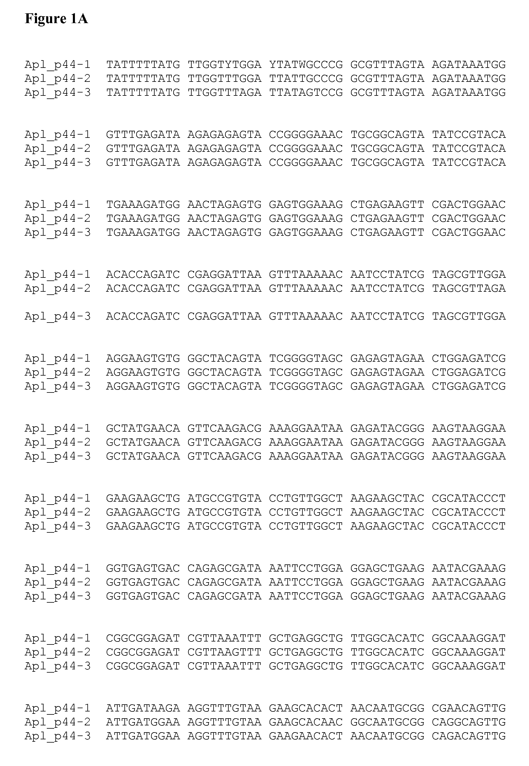
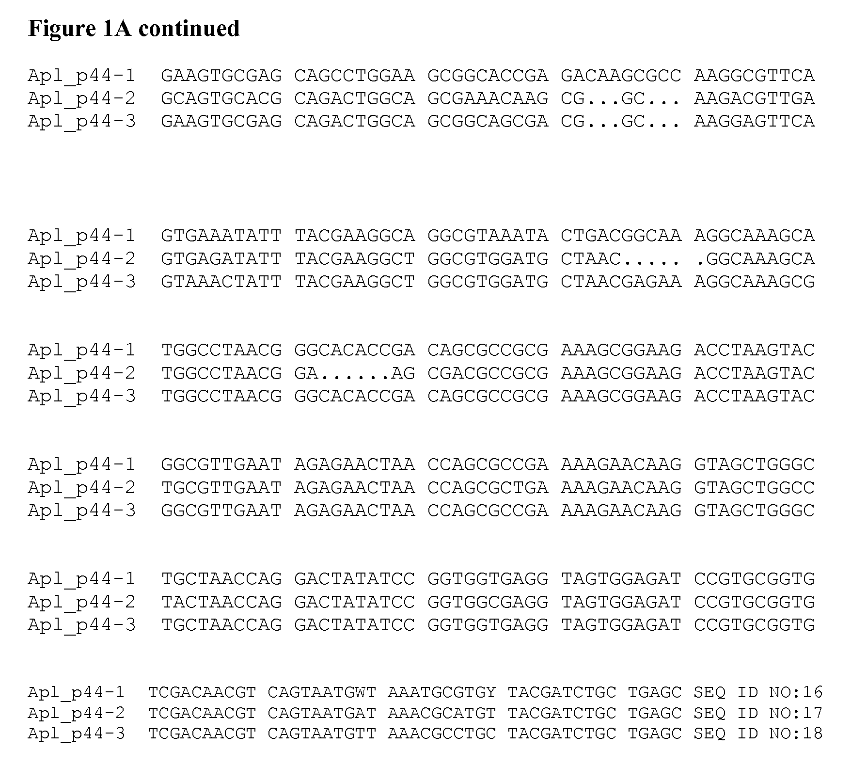
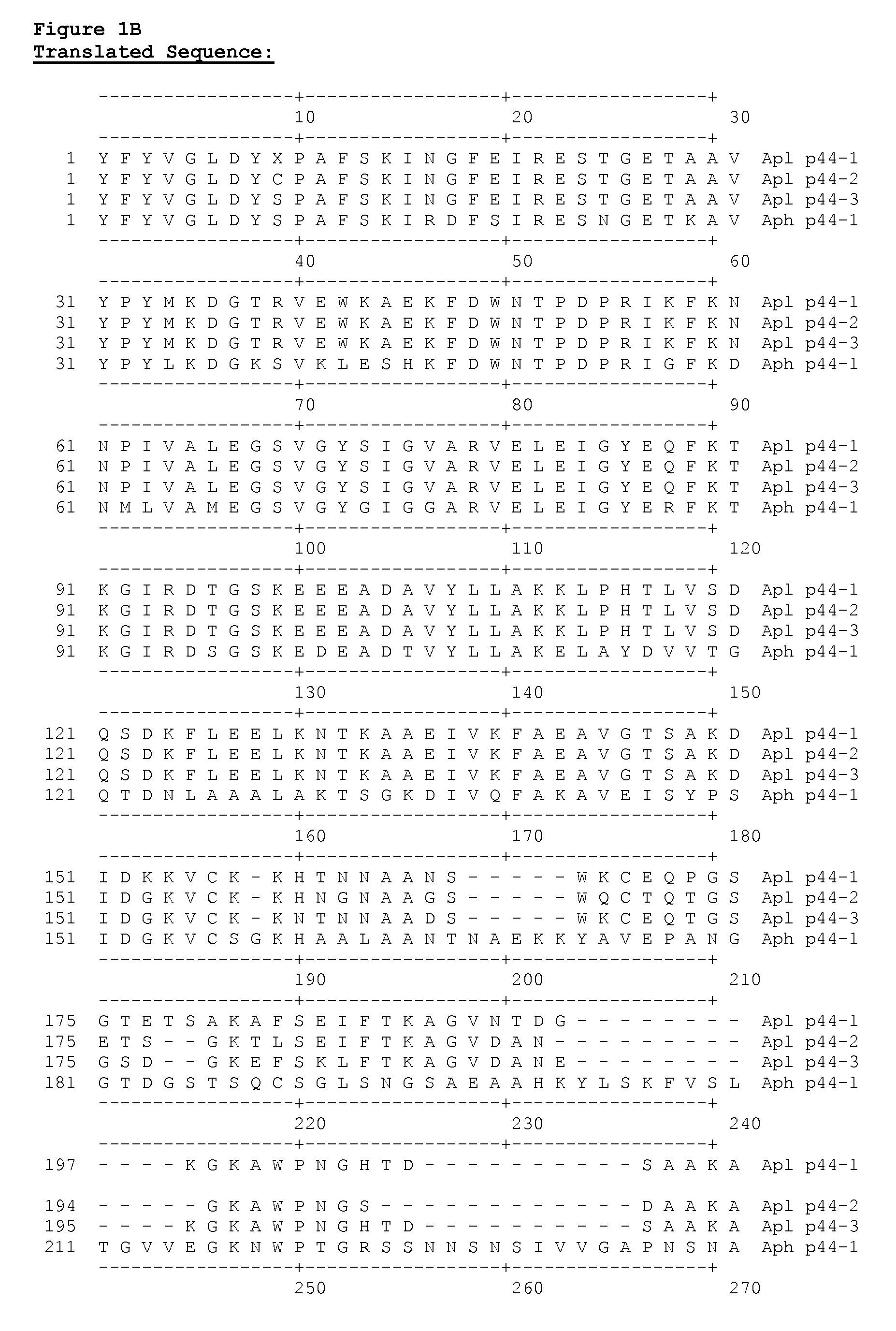
[0080]Degenerate primer-based PCR on a conventional thermocycler was used to amplify a polynucleotide with a length of approximately 800 nucleotides from an Apl p44 gene according to standard protocols (Platinum® Taq, Invitrogen). PCR products were cloned, sequenced, and analyzed relative to those reported for other species of Anaplasma. FIG. 1A shows ...
example 2
Detection of Apl by Real Time PCR Assay
[0081]A real-time PCR assay was developed to detect an Apl p44 polynucleotide from genomic DNA. Sample types for analysis included canine whole blood, as well as nymph and adult ticks. Primers and hybridization probes were selected to be specific for an Apl p44 gene and did not amplify the p44 gene of Aph. Sequences of the primers and probes are shown below:
[0082]
Apl p44 forward primer:5′ CCGGCGTTTAGTAAGATAAATG 3′(SEQ ID NO: 6)Apl p44 reverse primer:5′ GCAAATTTAACGATCTCCGCC 3′(SEQ ID NO: 7)Apl p44 probe 1129-FITC:5′ ACAGTATCGGGGTAGCGAGAGTAGAA 3′(SEQ ID NO: 8)Apl p44 probe 1183-LC670:5′ GGAGATCGGCTATGAACAGTTCAAGAC 3′(SEQ ID NO: 9)
[0083]These were synthesized by a commercial vendor. The real-time PCR was optimized for the Roche LightCycler® 480 using Roche reagents (Genotyping Master Mix #04707524001). Primers were used at a concentration of 0.3 μM for the forward primer and 0.6 μM for the reverse primer. Both probes were used at a concentration ...
example 3
Detection of Apl by an Anti-Species, Indirect ELISA
[0084]A synthetic peptide derived from the p44 gene of Apl was tested in an ELISA format to determine serological reactivity in dogs from an area with a high burden of Rhipicephalus ticks and high seroprevalence for E. canis. These geographic areas have been shown to have relatively high levels of A. platys infections and an absence of A. phagocytophilum infections in dogs by PCR. The peptide sequence is shown below:
[0085]
(SEQ ID NO: 15)Cys-Lys-Asp-Gly-Thr-Arg-Val-Glu-Trp-Lys-Ala-Glu-Lys-Phe-Asp-Trp-Asn-Thr-Pro-Asp-Pro-Arg-Ile.
[0086]An alternate peptide sequence comprises:
[0087]
(SEQ ID NO: 14)Cys-Lys-Asp-Gly-Thr-Arg-Val-Glu-Trp-Lys-Ala-Glu-Lys-Phe-Asp-Trp-Asn-Thr-Pro-Asp-Pro-Arg-Ile-Lys-Phe-Lys-Asn
[0088]The synthetic peptide of SEQ ID NO: 15 was solubilized in DMSO and coated on Immulon 2Hb plates (Thermo Electron Corporation #3455) at a concentration of 0.25 μg / ml in 50 mM Tris, pH 7.5 overnight at room temperature. The plates were...
PUM
| Property | Measurement | Unit |
|---|---|---|
| temperatures | aaaaa | aaaaa |
| temperatures | aaaaa | aaaaa |
| temperature | aaaaa | aaaaa |
Abstract
Description
Claims
Application Information
 - R&D
- Intellectual Property
- Life Sciences
- Materials
- Tech Scout
- Unparalleled Data Quality
- Higher Quality Content
- 60% Fewer Hallucinations
Browse by: Latest US Patents, China's latest patents, Technical Efficacy Thesaurus, Application Domain, Technology Topic, Popular Technical Reports.
© 2025 PatSnap. All rights reserved.Legal|Privacy policy|Modern Slavery Act Transparency Statement|Sitemap|About US| Contact US: help@patsnap.com


