Method and medical imaging system for compensating for patient motion

a technology of medical imaging and patient motion, applied in the field of medical imaging system for compensating patient motion, to achieve the effect of reducing the time outlay of the operator and improving the image results

Inactive Publication Date: 2009-12-08
SIEMENS HEALTHCARE GMBH
View PDF18 Cites 9 Cited by
  • Summary
  • Abstract
  • Description
  • Claims
  • Application Information

AI Technical Summary

Benefits of technology

[0014]Based on this prior art, an object of the present invention is to specify a method and an associated imaging system to compensate for patient motion in series recordings in medical imaging, with which patient motion can be compensated for during image recording without time-consuming user interaction, the method being such that it can be implemented in real time. The method and the associated imaging system are intended in particular to improve image results for digital subtraction angiography and road mapping with the smallest possible time outlay for the operator.
[0020]Unlike most previously known methods for motion correction, the present method does not require interaction with the operator. The previous methods for retrospective image processing can operate in principle only approximately. Major motion can barely be corrected with these methods and minor motion only approximately. The proposed method operates with great precision in particular even with major motion, so that the need for multiple recording of a mask image is dispensed with. This saves time and contrast agent and reduces the X-ray dose in the case of X-ray recordings. The present method also allows sedation or anesthesia of the patient just for the purposes of minimizing motion artifacts to be dispensed with in certain cases.
[0023]To compensate for the differences detected, it is also possible to change the image content of the recorded images geometrically. This relates in particular to a rotation of the image content about an axis perpendicular to the image plane and translation in the two degrees of freedom of translation of the two-dimensional image recording. It is also possible to scale the image. Depending on the type of patient motion a combination of the two compensation techniques, i.e. changing the geometric relationships of the imaging system and geometric adjustment of the image content, can be advantageous.
[0024]Naturally with the present method retrospective image processing methods can also be used after the at least approximate compensation for motion, to improve the image results still further. Approximate compensation by changing the geometric relationships of the imaging system can thereby be used to compensate for gross motion, while minor residual errors are eliminated by retrospective image processing.

Problems solved by technology

The latter is only possible when recording the 3D image data set with the same imaging system.

Method used


Image

  • Method and medical imaging system for compensating for patient motion
  • Method and medical imaging system for compensating for patient motion
  • Method and medical imaging system for compensating for patient motion

Examples


Embodiment Construction

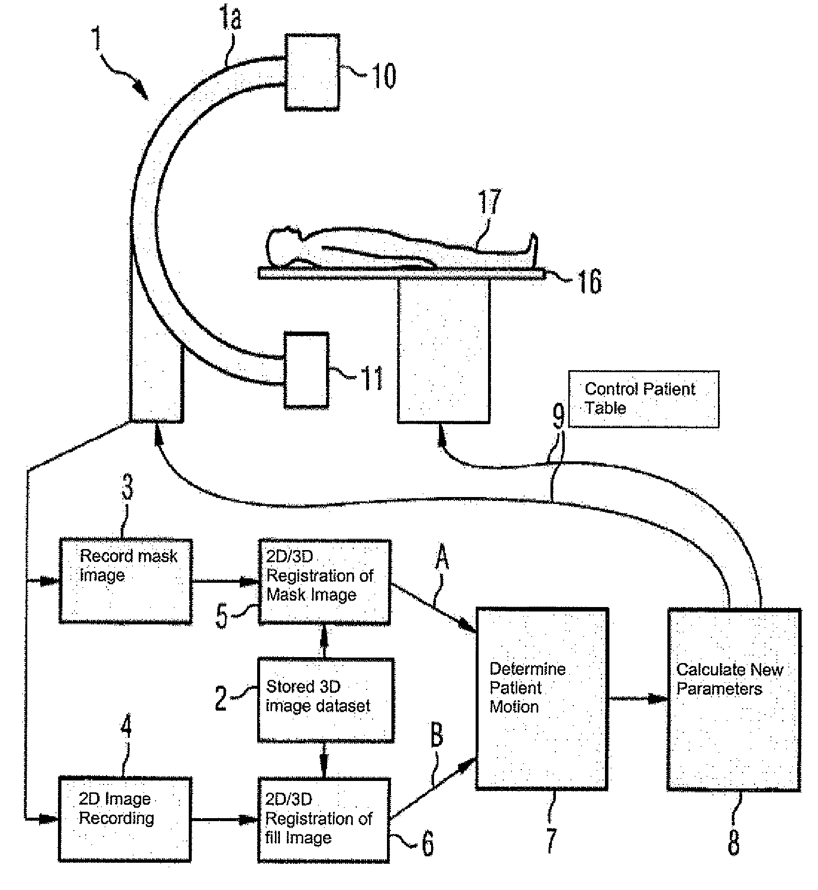
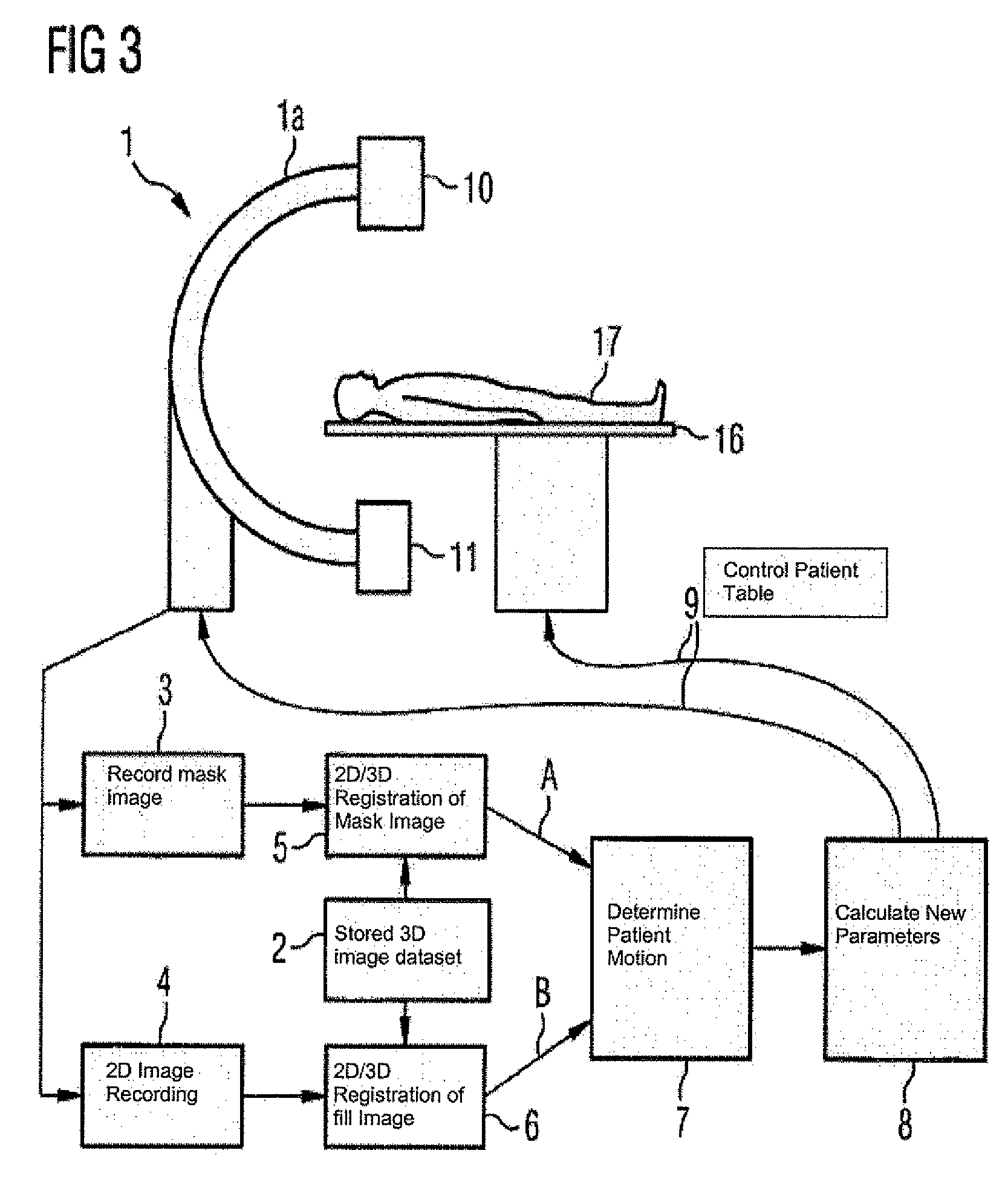
[0030]The present method is described below with reference to an X-ray angiography unit for neuroradiology applications. The method can of course also be used in other fields, in which digital subtraction angiography and / or road mapping are used. The present method can also be used with other techniques for medical imaging, in which series recordings have to be taken and related to each other.

[0031]In the example below the embodiments are restricted to the instance of the correction of head motion of a patient. As the head can be considered approximately to be a rigid element, the motion correction is restricted to the six degrees of freedom of translation and rotation of a rigid element in the three-dimensional space.

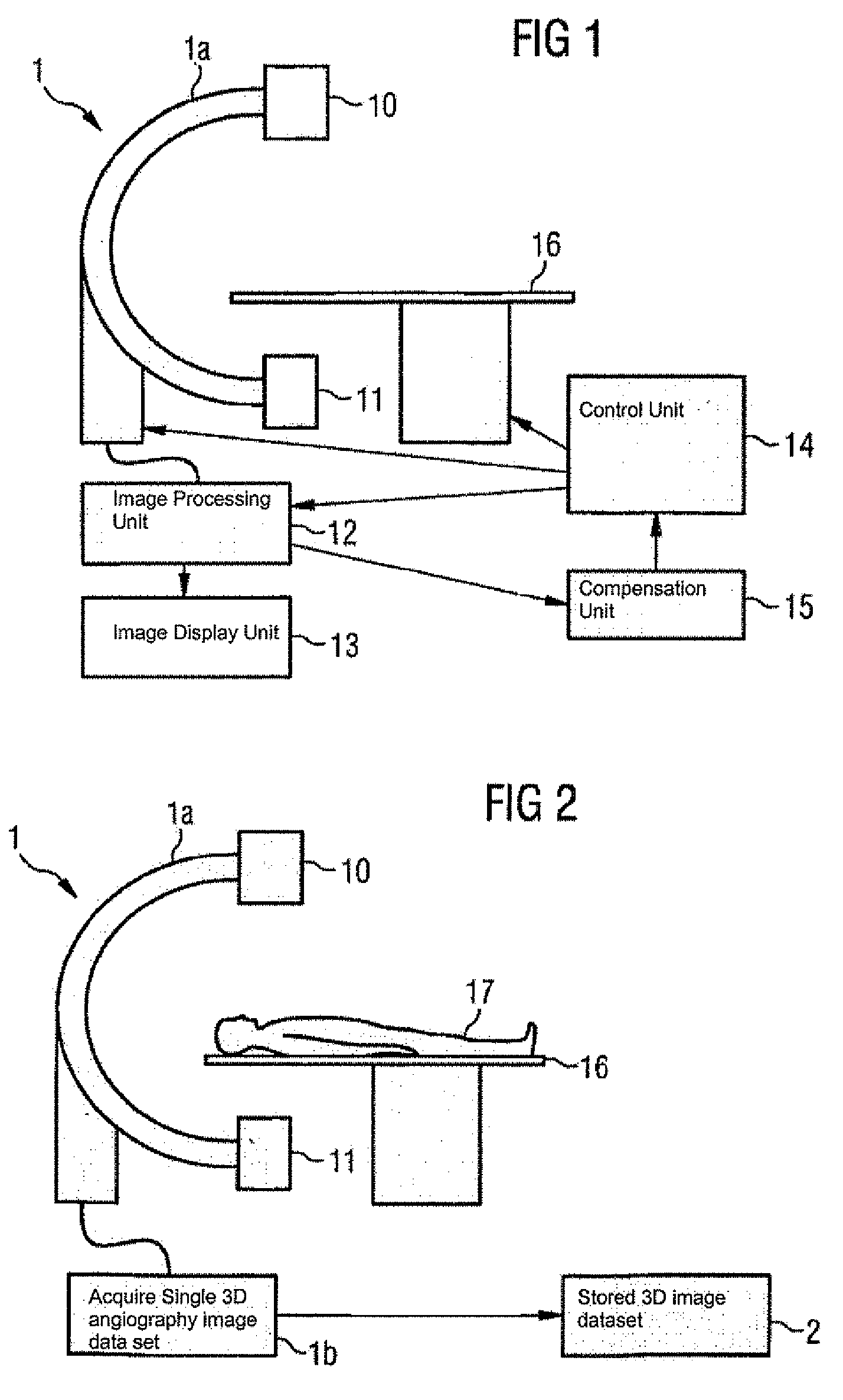
[0032]A neuroradiology X-ray angiography unit 1 is used for image recording, as shown schematically in FIG. 1. The X-ray angiography unit 1 includes a C-arm 1a that can be rotated about two axes, to which an X-ray tube 10 and a detector 11 opposite the X-ray tube are a...


PUM


Abstract

Disclosed herein is a method, and system (1) for implementing same, to compensate for patient motion in series recordings in medical imaging, in which a plurality of images of an examination area of a patient (17) are recorded at time intervals with an imaging system (1) and related to each other. Before the start of the series recordings a 3D image data set is recorded by a 3D recording of the examination area, which establishes a reference system. A first spatial position of the examination area in the reference system is obtained by recording a first image of the series recordings and then registering it or by calculating it. Each further image of the series recordings is registered immediately after recording to obtain the current spatial position of the examination area. Differences in spatial position are compensated for at least approximately by changing geometric relationships of the imaging system (1).

Description

CROSS REFERENCE TO RELATED APPLICATIONS[0001]This application claims priority to the German application No. 10 2004 004 603.4, filed Jan. 29, 2004 which is incorporated by reference herein in its entirety.FIELD OF INVENTION[0002]The present invention relates to a method to compensate for patient motion in series recordings in medical imaging, with which a plurality of images of an examination area of a patient are recorded at time intervals using an imaging system and related to each other, in particular to compensate for motion in the case of digital subtraction angiography or the pathfinder technique. The invention also relates to an imaging system with a radiation source, detector and patient table as well as a control, image processing and image display unit, which is configured to implement the method.BACKGROUND OF INVENTION[0003]In one of the main fields of application of the present method, namely the field of digital subtraction angiography, blood vessels in the human body a...

Claims


Application Information

Patent Timeline
Patent Type & Authority Patents(United States)
IPC IPC(8): A61B5/05A61B6/00G06T7/00G06T7/20
CPCA61B6/481A61B6/504A61B6/527G06T7/0038G06T7/004G06T7/20G06T2207/30004A61B6/5288A61B6/5264G06T7/38G06T7/70
Inventor BOESE, JANHEIGL, BENNORAHN, NORBERT
Owner SIEMENS HEALTHCARE GMBH
Why Eureka
Try Eureka