Endoscopic bite block for use with cannula

a technology of endoscopic instruments and cannulas, which is applied in the field of endoscopic bite blocks, can solve the problems of patient biting on endoscopic instruments, certain well-known risks related to patient respiration, hypoventilation, and oxygen desaturation

Inactive Publication Date: 2011-10-04
ETHICON ENDO SURGERY INC
View PDF59 Cites 11 Cited by
  • Summary
  • Abstract
  • Description
  • Claims
  • Application Information

AI Technical Summary

Problems solved by technology

The bite block or mouthguard essentially maintains the patient's mouth in the open position, providing an opening through which the endoscope can be passed, and prevents the patient from biting down on the endoscopic instruments, which are often quite expensive.
In the case when the particular sedation drugs are respiratory depressants, there exist certain well-known risks related to patient respiration, including hypoventilation, oxygen desaturation, and apnea.
Difficulties sometimes arise, however, when simultaneously managing the scope, delivering supplementary oxygen, and sampling respiratory gasses via the oral cavity.
Unfortunately, this method would require that the patient inhale and exhale only through the nasal passages for the duration of the procedure; in a real-world scenario, however, this is not the case.
It indeed requires little imagination to see that accommodating all three activities simultaneously through the oral cavity with instruments not designed to be used together would prove troublesome.
While focusing on the endoscope, an oral-nasal cannula is rather easily bumped and relocated during the maneuvering of the scope, leaving its oral ports situated too far from the oral cavity and occasionally causing bruising internal to the nasal passages.
The consequence is decreased effectiveness in the administration of supplementary oxygen and sampling of respiratory gasses, which in turn may compromise patient safety.
This requires that the finger be inserted at least to the depth of the end of the bite block, which may cause the bite block to move around.
This adds to the risk that, during all of the jostling of the bite block associated with the maneuvering of the endoscope and insertion of a finger, the oral ports of the cannula may be unintentionally relocated away from the oral cavity.

Method used


Image

  • Endoscopic bite block for use with cannula
  • Endoscopic bite block for use with cannula
  • Endoscopic bite block for use with cannula

Examples


Embodiment Construction

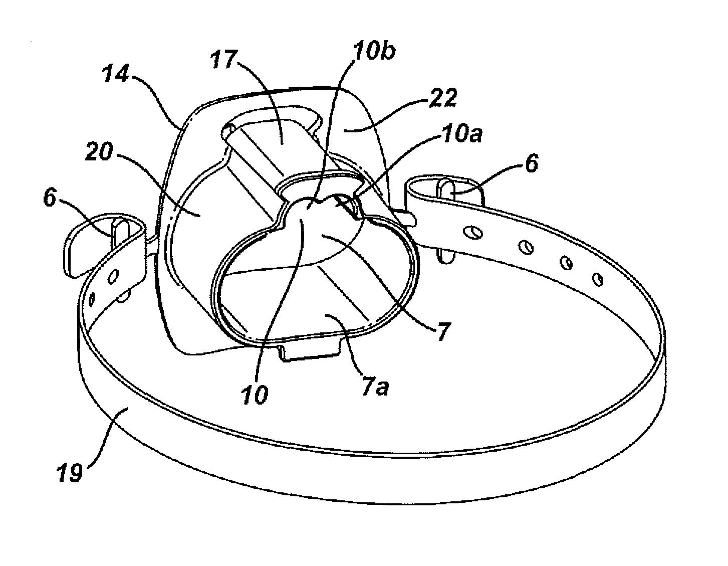
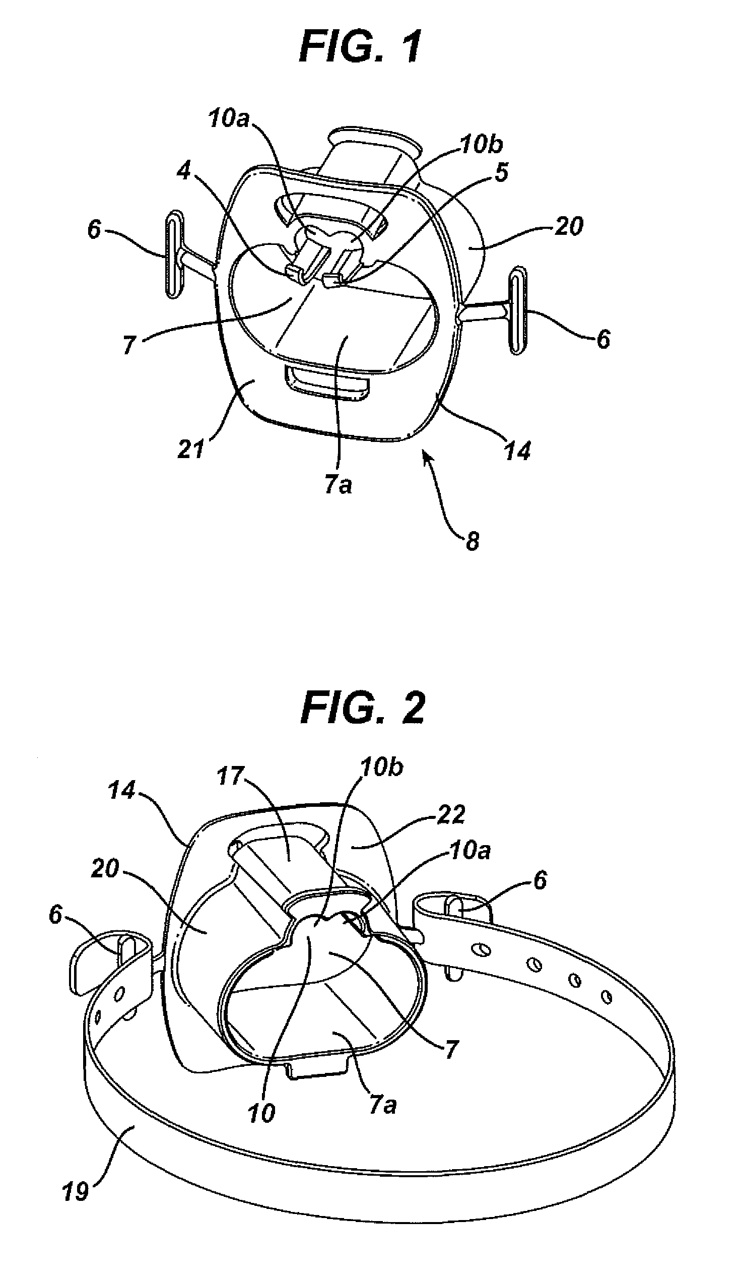
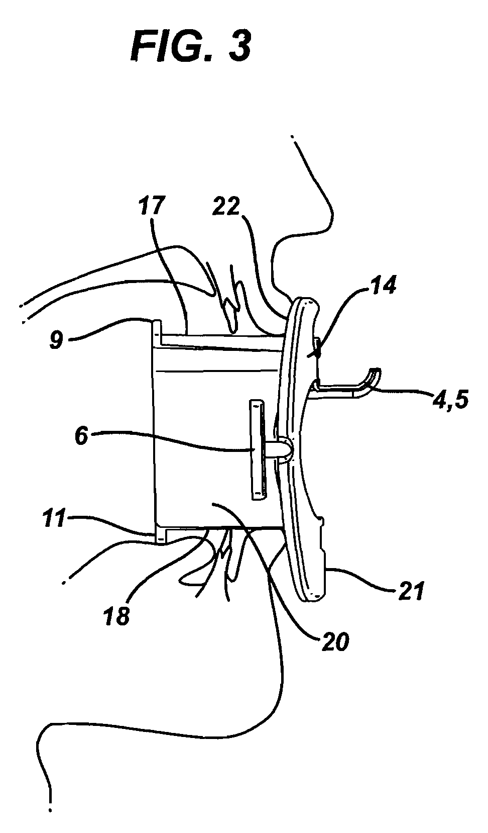
[0016]Referring to FIG. 1 and FIG. 2, the bite block 8 of the present invention consists of a generally elliptical cylindrical main body 20, having a proximal end, which sits outside of a patient's mouth, and a distal end, which sits inside a patient's mouth. Main body 20 surrounds main oral passage lumen or channel 7, which is sized to allow for passage of an endoscope and ventilation of the patient. Oral passage 7 is defined by a lower surface 7a and an upper surface 7b (FIG. 5). Upper surface 7b further defines an opening for internal gas channel or lumen 10 (FIG. 5). Integral to the proximal end of main body 20 is flange 14, which sits outside of a patient's lips and serves both to locate bite block 8 relative to the patient's mouth and protect the patient's lips and teeth from an endoscope. Flange 14 is integral to main body 20 at distal surface 22. Attached at each side of flange 14 is strap attachment wing 6 for strap 19 that goes around the patient's head and helps secure bi...


PUM


Abstract

A bite block that is inserted into a patient's mouth during an endoscopic diagnostic or surgical procedure that has a channel for receiving an endoscope or other surgical instrument through the patient's mouth and additional channels transmitting a gas to the patient and transmitting expired gas from the patient.

Description

CROSS REFERENCE TO RELATED APPLICATIONS[0001]The present application claims the priority benefit of U.S. provisional patent application Ser. No. 60 / 941,707, filed on Jun. 4, 2007, the contents of which are incorporated by reference in their entirety herein.FIELD OF THE INVENTION[0002]The present invention relates, in general, to bite blocks for use in endoscopic surgical procedures, and in particular, to endoscopic bite blocks for use in procedures involving sedation and analgesia systems.BACKGROUND OF THE INVENTION[0003]During some medical procedures, specifically endoscopic procedures, it is necessary to insert medical instruments, such as an endoscope, into the mouth and down the trachea or esophagus of a patient. It is common to use in such procedures a bite block or mouthguard to protect both the patient's mouth from the endoscope and the endoscope from the patient's mouth. The bite block or mouthguard essentially maintains the patient's mouth in the open position, providing an...

Claims


Application Information

Patent Timeline
Patent Type & Authority Patents(United States)
IPC IPC(8): A61C5/14
CPCA61M16/0666A61M16/0493A61M16/0683A61B1/00154A61B1/24A61M16/0497A61M2202/0208A61M2230/432A61M16/085
Inventor REYNOLDS, II, DONALD L.STEPHENS, RANDY R.FLAKER, RICHARD W.
Owner ETHICON ENDO SURGERY INC
Why Eureka
Try Eureka