Method and apparatus for preprocessing ultrasound imaging

a preprocessing and ultrasound technology, applied in the field of preprocessing ultrasound imaging, can solve the problems of limiting its application, difficulty in therapy, and the inability of conventional medical imaging equipment to offer a two-dimensional image of the inside human organ, and achieve the effect of effectively removing speckle noise and effectively protecting the edg

Active Publication Date: 2011-11-15
SHENZHEN MINDRAY BIO MEDICAL ELECTRONICS CO LTD
View PDF19 Cites 0 Cited by
  • Summary
  • Abstract
  • Description
  • Claims
  • Application Information

AI Technical Summary

Benefits of technology

[0021]According to the method for preprocessing ultrasound imaging adopted in the technical solution of the present invention, on the object's surface in the volume data, the branches of the tangent tree distribute along the surface as much as possible without crossing or breaking away from the surface, which can protect the edge effectively. At the same time, the speckle noise can be eliminated effectively by calculating the mean value of plurality of nodes distributed along the surface, thus achieving the purpose of smoothing. So, this algorithm can give consideration to both smoothness and details.

Problems solved by technology

A conventional medical imaging equipment can only offer a two-dimensional image of the inside human organs.
A doctor can empirically estimate the size and shape of nidus from a plurality of 2D images so as to “conceive” a three-dimensional geometrical relationship between the nidus and its surrounding tissues, which brings difficulty to therapy.
However, speckle noise specific to the ultrasound image has become an important factor influencing the imaging effect, thus limiting its application.
In forming the ultrasound image, reflection echoes from the reflection points whose space is smaller than a wavelength interfere with each other so as to produce the speckle noise, which will blur the image features and disturb observation and analysis.
If such a volume data not being processed is used in 3D imaging directly, the resulting visual effect will be unacceptable.
The shortcoming of the above-mentioned technology is that it can not be used in 3D application directly.
Even though it is extended to 3D application and the size of the template is adjusted, either the noise is significant or the imaging result becomes blurry, unable to smooth the imaging data without compromising important details.
Therefore, the prior techniques are not adapted for practical application.

Method used


Image

  • Method and apparatus for preprocessing ultrasound imaging
  • Method and apparatus for preprocessing ultrasound imaging
  • Method and apparatus for preprocessing ultrasound imaging

Examples


Embodiment Construction

1. Method for Preprocessing an Ultrasound Image

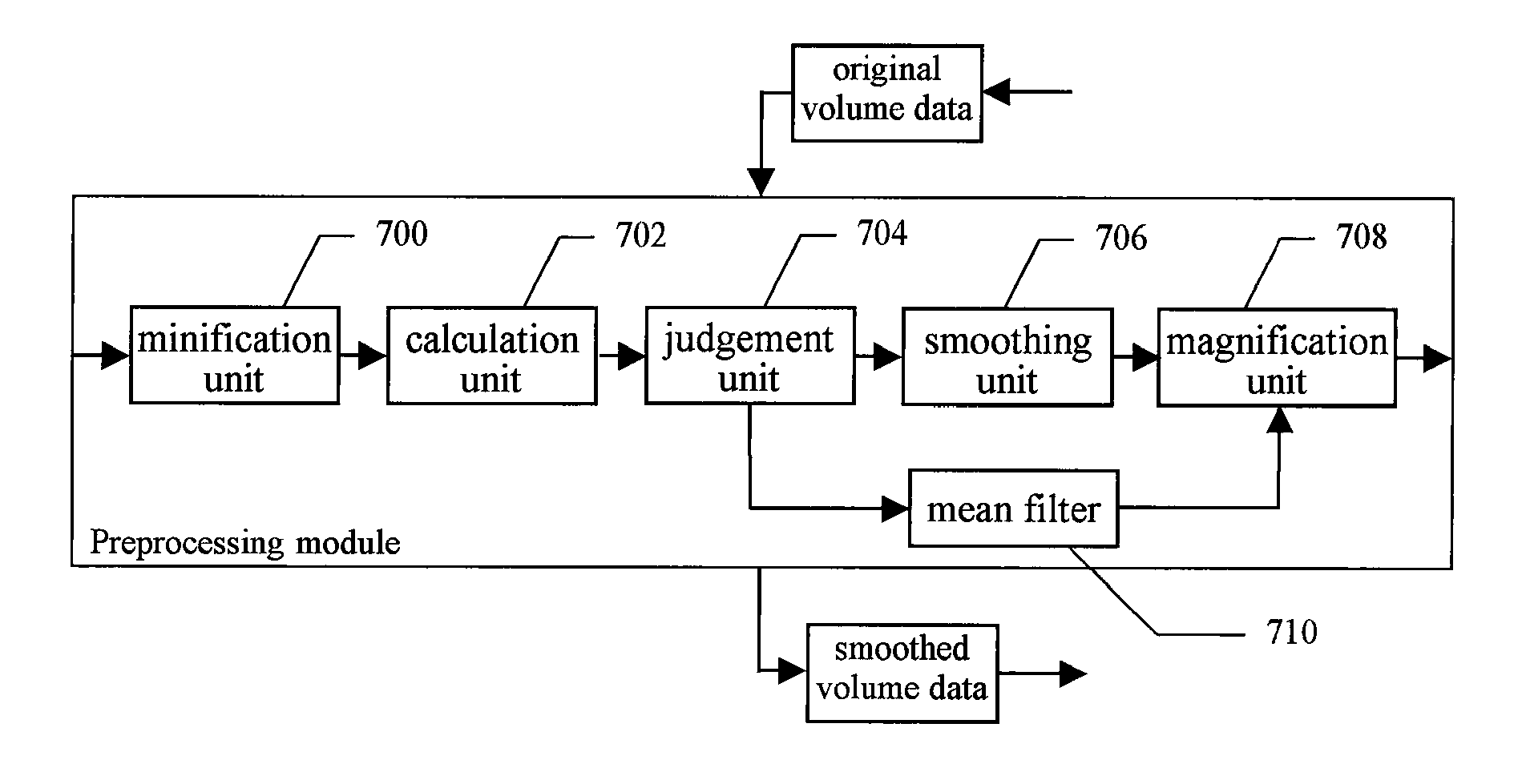
[0030]The method for preprocessing an ultrasound image according to the embodiment of the present invention is described as follows, taking a 3D ultrasound image as an example.

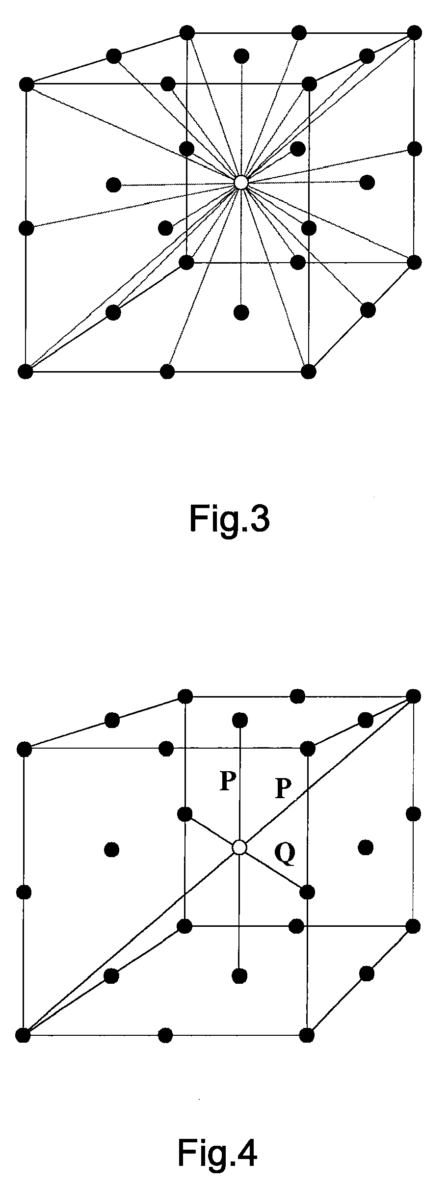
[0031]Put it briefly, the method for preprocessing ultrasound imaging according to the embodiment of the present invention first minifies volume data before digital scanning conversion (DSC) and then performs the core algorithm and finally magnifies the data to its original size. In the core algorithm process, a cubic neighborhood is first determined for each voxel, which centers around that voxel and has a “radius” of R (“radius” mentioned herein refers to a distance from the center of the cube to its surface, i.e., the length of the cube's side equals to 2R+1). Subsequently, 13 line segments centering at the current voxel are examined in respect of uniformity. The most uniform N directional lines are then selected to generate 2N optimal directions. After each vo...


PUM


Abstract

The present invention provides a method and module for preprocessing ultrasound imaging. The method comprises a calculation step for constructing a multivalue vector field and a smoothing step for smoothing the whole volume data. The method further comprises a judgement step and minification and magnification steps. The module includes a calculation unit, a smoothing unit, a judgement unit, a minification unit and a magnification unit. According to the method for preprocessing ultrasound imaging, speckle noise can be eliminated effectively by calculating a mean value of a plurality of nodes distributed over the surface, so as to implement the smoothing. Therefore, this method is capable of smoothing data without compromising details.

Description

STATEMENT OF RELATED APPLICATION[0001]The present application claims priority of the Chinese Patent Application No. 200610146989, entitled “Method and Apparatus for Preprocessing Ultrasound Imaging”, filed on Nov. 28, 2006, which is incorporated herein by reference in its entirety.FIELD OF THE INVENTION[0002]The present invention relates to a method and apparatus for processing an image signal, and specifically to a method for preprocessing ultrasound imaging and an apparatus for implementing the same.BACKGROUND OF THE INVENTION[0003]A conventional medical imaging equipment can only offer a two-dimensional image of the inside human organs. A doctor can empirically estimate the size and shape of nidus from a plurality of 2D images so as to “conceive” a three-dimensional geometrical relationship between the nidus and its surrounding tissues, which brings difficulty to therapy. A 3D visual technology can reconstruct a 3D image from a series of 2D images and display it on a terminal. Th...

Claims


Application Information

Patent Timeline
Patent Type & Authority Patents(United States)
IPC IPC(8): G06K9/00G06K9/36
CPCG06T15/08G06T5/002G06T5/20G01S15/8993G06T2200/04G06T2207/10132G06T2207/20192
Inventor TIAN, YONGYAO, BINHU, QINJUN
Owner SHENZHEN MINDRAY BIO MEDICAL ELECTRONICS CO LTD
Why Eureka
Try Eureka