Method and system for 3D cardiac motion estimation from single scan of C-arm angiography

a single scan and c-arm technology, applied in the field of cardiac imaging, can solve the problems of difficult to ask a patient to hold his or her breath for such a long period, the accuracy of the reconstruction 3d image remains a question, and it is difficult to estimate the motion of the cardiac muscl

Active Publication Date: 2014-12-30
SIEMENS HEALTHCARE GMBH
View PDF20 Cites 12 Cited by
  • Summary
  • Abstract
  • Description
  • Claims
  • Application Information

AI Technical Summary

Problems solved by technology

Heart disease affects a large number of people globally and has become the leading cause of death in the United States.
However, due to cardiac motion, the reconstructed 3D image is typically blurred due to averaging from all projections belonging to different cardiac phases.
However, the accuracy of the image after transformation remains a question.
In real clinical practice, it is difficult to ask a patient to hold his or her breath for such a long period, especially for a severely ill patient or a patient under general anesthesia.
In addition, multiple sweeps consume more contrast agent, which often causes side effects (e.g., allergy or renal insufficiency), and expose patients to more radiation.
If only a single sweep is applied, there are a limited number of projection images available for each cardiac phase, which results in reconstruction artifacts due to missing data or residual motion.

Method used


Image

  • Method and system for 3D cardiac motion estimation from single scan of C-arm angiography
  • Method and system for 3D cardiac motion estimation from single scan of C-arm angiography
  • Method and system for 3D cardiac motion estimation from single scan of C-arm angiography

Examples


Embodiment Construction

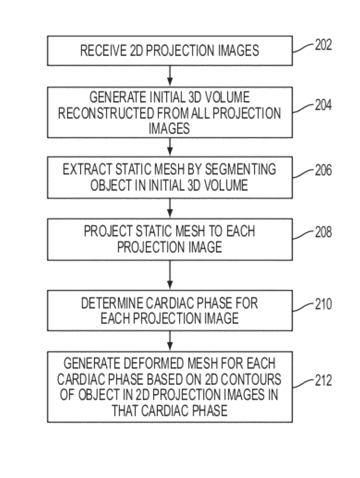
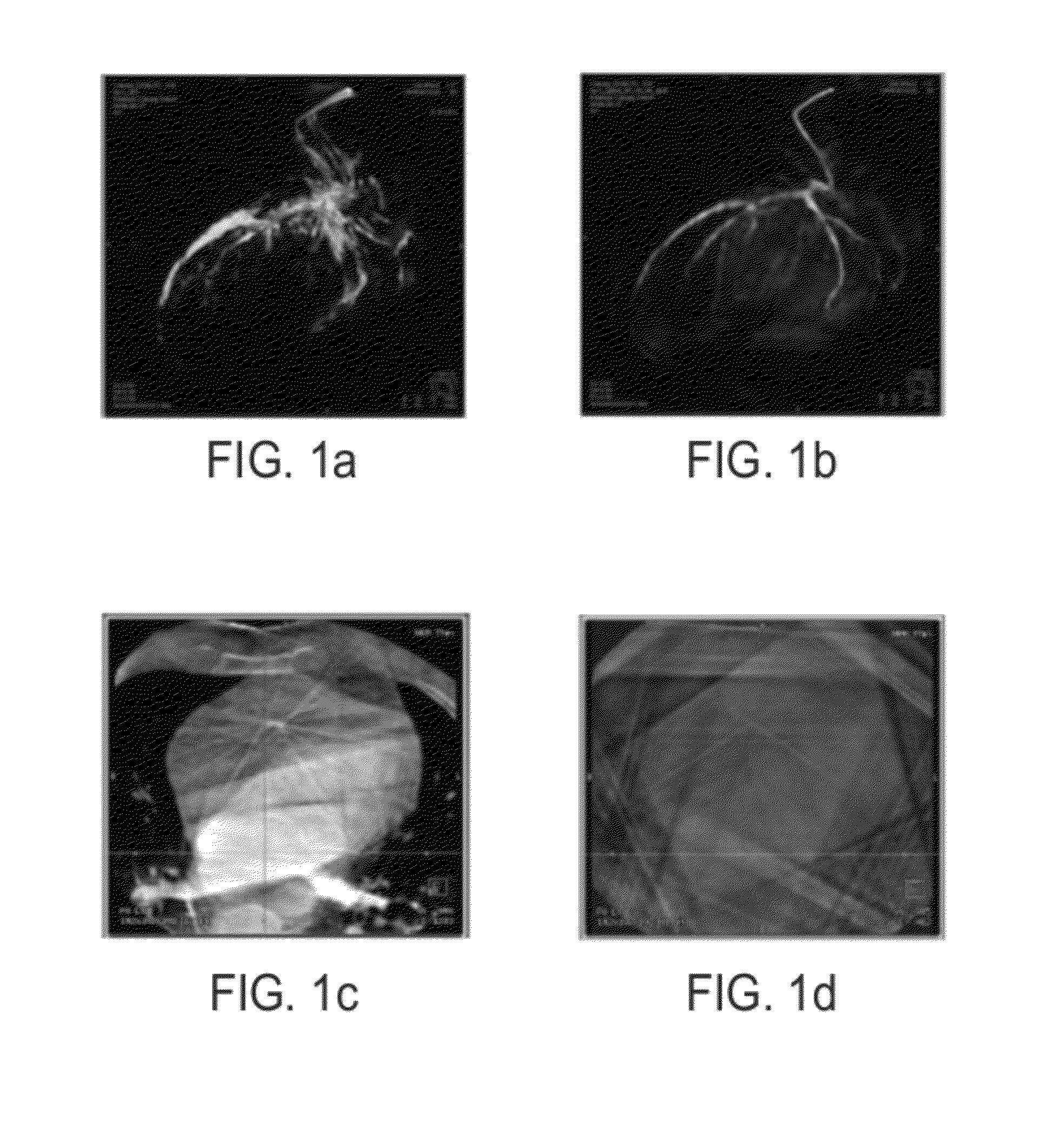
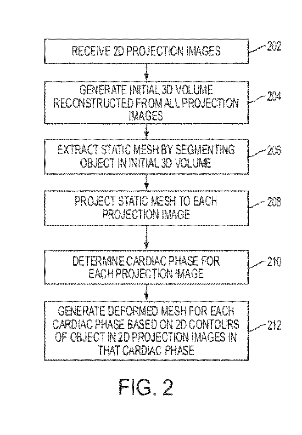
[0020]The present invention is directed to a method and system for three-dimensional (3D) cardiac motion estimation from a single scan of C-arm angiography. Embodiments of the present invention are described herein to give a visual understanding of the 3D cardiac motion estimation method. A digital image is often composed of digital representations of one or more objects (or shapes). The digital representation of an object is often described herein in terms of identifying and manipulating the objects. Such manipulations are virtual manipulations accomplished in the memory or other circuitry / hardware of a computer system. Accordingly, it is to be understood that embodiments of the present invention may be performed within a computer system using data stored within the computer system.

[0021]Using a C-arm image acquisition system, it is possible to generate a 3D reconstructed computed tomography (CT) image by reconstructing a 3D image from 2D projections. However, due to cardiac motion...


PUM


Abstract

A method and system for estimating 3D cardiac motion from a single C-arm angiography scan is disclosed. An initial 3D volume is reconstructed from a plurality of 2D projection images acquired in a single C-arm scan. A static mesh is extracted by segmenting an object in the initial 3D volume. The static mesh is projected to each of the 2D projection images. A cardiac phase is determined for each of the 2D projection images. A deformed mesh is generated for each of a plurality of cardiac phases based on a 2D contour of the object and the projected mesh in each of the 2D projection images of that cardiac phase.

Description

[0001]This application claims the benefit of U.S. Provisional Application No. 61 / 434,558, filed Jan. 20, 2011, the disclosure of which is herein incorporated by reference.BACKGROUND OF THE INVENTION[0002]The present invention relates to cardiac imaging, and more particularly, to estimating 3D cardiac motion from a single scan of C-arm angiography.[0003]Heart disease affects a large number of people globally and has become the leading cause of death in the United States. The estimation of cardiac motion and deformation is an area of concern in medical image analysis, as such cardiac motion and deformation has important clinical implications for the viability of the heart muscle. Various studies have attempted to address estimating cardiac motion and deformation in different imaging modalities, including cardiac computed tomography (CT), ultrasound, and magnetic resonance imaging (MRI).[0004]The recent development of the C-arm image acquisition system provides greater flexibility by e...

Claims


Application Information

Patent Timeline
Patent Type & Authority Patents(United States)
IPC IPC(8): G06K9/00G06T7/00
CPCG06T7/0083G06T2207/10081G06T7/0089G06T2207/30048G06T7/12G06T7/149
Inventor CHEN, MINGQINGZHENG, YEFENGFUNKA-LEA, GARETHLAURITSCH, GUENTERBOESE, JANCOMANICIU, DORIN
Owner SIEMENS HEALTHCARE GMBH
Why Eureka
Try Eureka