
Proteome-wide quantification of small molecule binding to cellular target proteins

a small molecule and protein technology, applied in the field of proteome-wide quantification of small molecule binding to cellular target proteins, can solve the problems of inability to infer the affinities of the inhibitor towards its cellular binding partner from the ms data, the underrepresentation or the complete absence of potential targets from other enzyme classes in the screening format, and the inability to characterize the pharmacological effect of the inhibitor

Active Publication Date: 2015-02-24
MAX PLANCK GESELLSCHAFT ZUR FOERDERUNG DER WISSENSCHAFTEN EV
View PDF9 Cites 2 Cited by
  • Summary
  • Abstract
  • Description
  • Claims
  • Application Information

AI Technical Summary

Benefits of technology

[0009]The above objects are solved by the methods according to the present invention. By virtue of these methods, it is now possible to immediately determine the binding affinities of a target compound, such as an inhibitor, towards its cellular binding partners, either qualitatively or quantitatively, without having to resort to secondary in vitro binding or activity assays. The methods of the invention furthermore allow ranking a large number of cellular targets of an immobilized compound according to their affinities, and furthermore to assess their competitive binding properties with regard to a distinct inhibitor without further immobilization. This is exemplarily demonstrated herein on more than 100 cellular targets of kinase inhibitors. The new methods are broadly applicable and allow for the simple and rapid characterization of pharmacological and pharmacokinetic interactions and / or functions of any type of small molecules or other compound of interest in various biological systems.
[0025]According to any of the methods of the invention described herein, the target components in the analyte are preferably labelled with a detectable label. While the detectable label may be the same in each aliquot, the detectable label is preferably different in each aliquot, i.e., the target components in the first aliquot have a label which is different from the target components in the second aliquot and so on. This can be easily achieved by direct labelling of the aliquots with different detectable labels, or in the case of analytes, which are derived from tissue, tissue culture, cells, cell culture, or body fluids, by metabolic labelling in vivo or in culture using different detectable labels.

Problems solved by technology

The pharmaceutical industry today faces two fundamental challenges in its drug development to process, namely the identification of appropriate protein targets for disease intervention and the identification of high quality drug candidates which act specifically on these targets.
This approach undoubtedly provides valuable and quantitative data about drug selectivity, but has two major limitations: First, in addition to a significant part of the protein kinase complement (=the kinome), potential targets from other enzyme classes are underrepresented or missing altogether in these screening formats.
Secondly, and perhaps more importantly, the recombinant kinase collection included in a selectivity panel does not match the cellular profile of potential targets expressed in, for example, a cell system where the biological effects of a kinase inhibitor are investigated.
However, target component identification was limited in the sense that the affinities of the inhibitor towards its cellular binding partners could not be inferred from the MS data.
However, in practice, it is challenging to assess the whole target spectrum of a small molecule drug due to the fact that the required recombinant proteins are not all available or in vitro activity assays prove difficult to establish.
Although affinity purification techniques together with mass spectrometry (MS) have been successfully used to identify the interacting proteins of immobilized small molecule inhibitors, these previous proteomics approaches did not deliver information of cellular target affinities (Godl, K. et al., PNAS U.S.A., 100, 15434-9 (2003); Brehmer, D. et al., Cancer Res. 65, 379-82 (2005); Daub, H., Biochim. Biophys. Acta 1754, 183-90 (2005)).

Method used


Image

  • Proteome-wide quantification of small molecule binding to cellular target proteins
  • Proteome-wide quantification of small molecule binding to cellular target proteins
  • Proteome-wide quantification of small molecule binding to cellular target proteins

Examples


example 1

Compound Synthesis and Covalent Coupling

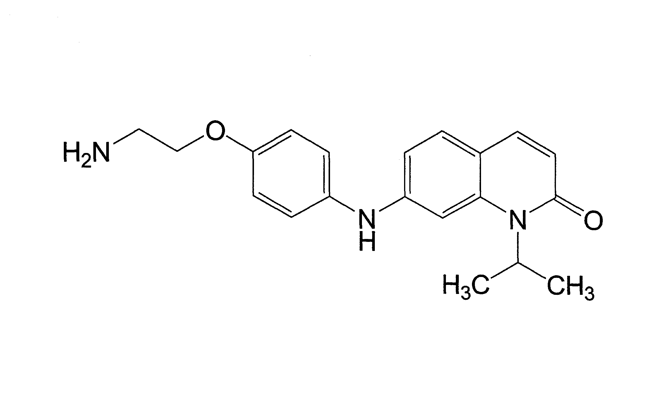
[0160]The kinase inhibitor V16742 was synthesised on the basis of described synthetic routes (Moloney, G. P. et al. A novel series of 2,5-substituted tryptamine derivatives as vascular 5HT1B / 1D receptor antagonists. J Med Chem 40, 2347-62 (1997). Barvian, M. et al. Pyrido[2,3-d]pyrimidin-7-one inhibitors of cyclin-dependent kinases. J Med Chem 43, 4606-16 (2000)). AX14596 and gefitinib were synthesized as described previously (Brehmer, D. et al. Cellular targets of gefitinib. Cancer Res 65, 379-382 (2005)). V16742 was coupled to epoxy-activated Sepharose beads at three different concentrations and the relative concentration of covalently immobilized inhibitor on the distinct resins was determined by spectrophotometry (here 1-, 5- and 25-fold of the lowest concentration). Two volumes of AX14596 at a concentration of 2.5 mM were coupled to one volume of epoxy-activated Sepharose beads (GE Healthcare) as previously described (Wissing, J. et al. P...

example 2

Cell Culture and In Vitro Association Experiments

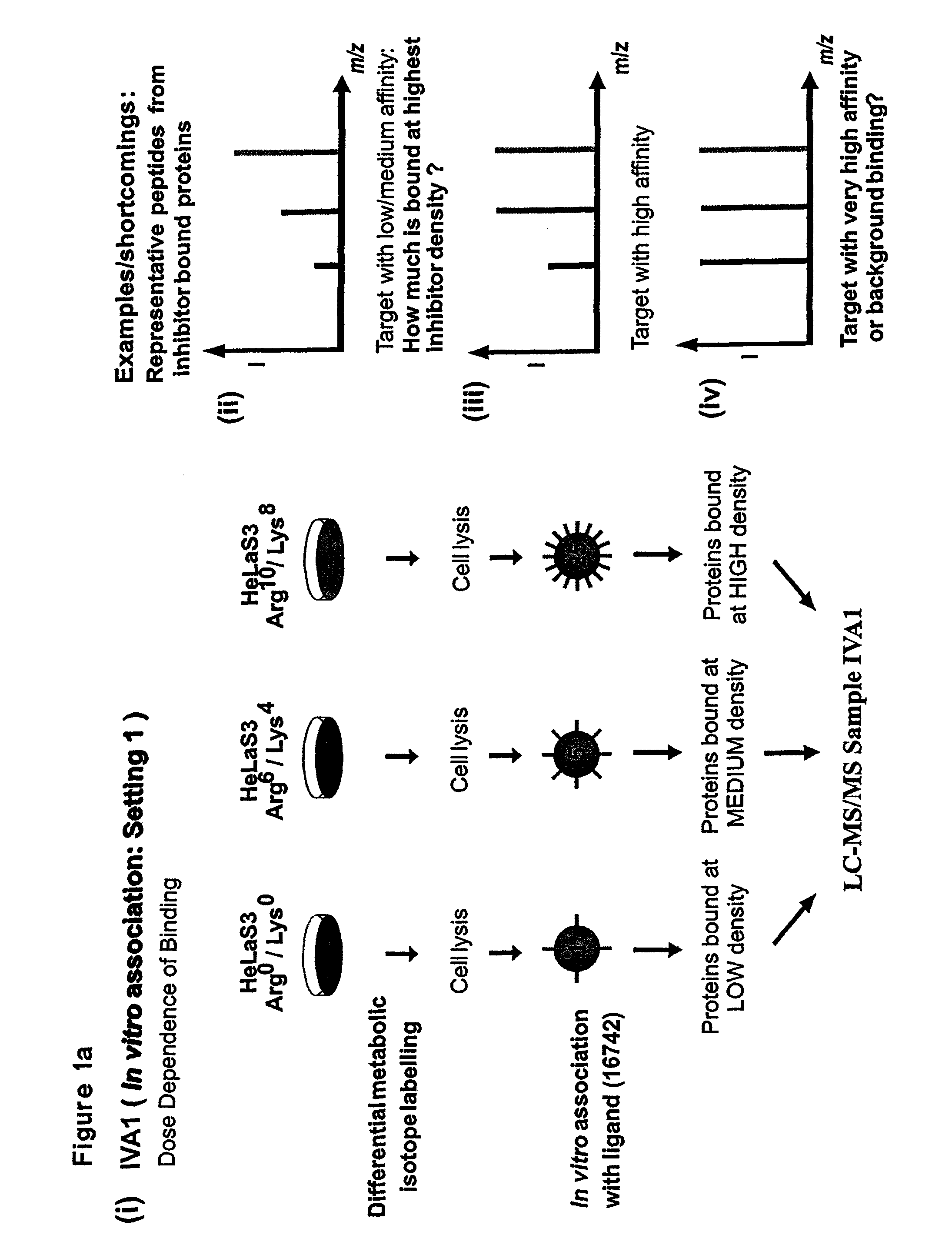
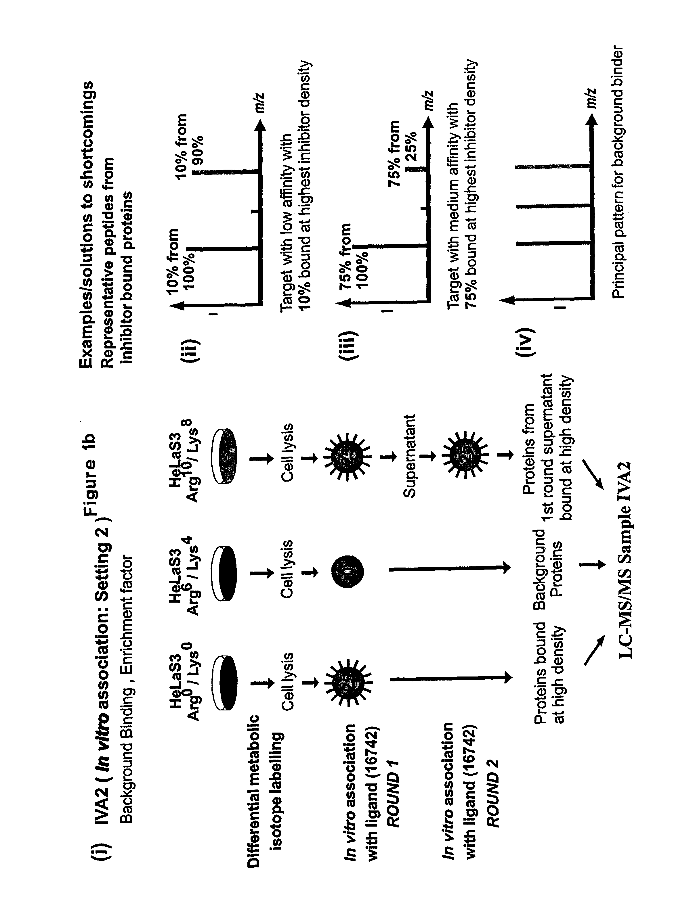
[0161]For stable isotope labelling with amino acids in cell culture (SILAC), HeLa S3 cells were grown in Dulbecco's modified Eagle's medium (DMEM) containing 10% dialysed fetal bovine serum (Invitrogen) and either unlabelled L-arginine)(Arg0) at 42 mg I−1 and L-lysine (Lys0) at 71 mg I−1 or equimolar amounts of the isotopic variants L-arginine-U-13C6 (Arg6) and L-lysine-2H4 (Lys4) or L-arginine-U-13C6—14N4 (Arg10) and L-lysine-13C6—15N2 (Lys8) (from Cambridge Isotope Laboratories).

[0162]Using the three populations of SILAC-labelled cells and the three different inhibitor resins, two parallel experiments termed as IVA1 (In Vitro Association, Setting 1) and IVA2 (In Vitro Association, Setting 2) were performed. For in vitro association with inhibitor affinity beads, differentially labeled HeLa cells were lysed in buffer containing 50 mM HEPES pH 7.5, 150 mM NaCl, 0.5% Triton X-100, 1 mM EDTA, 1 mM EGTA plus additives. After centrifugati...

example 3

IVA Experiments in the Presence of Competitor SB203580

[0163]Lysates from differentially SLAG-labeled cells were preincubated with increasing concentrations of the kinase inhibitor SB203580 (0 nM, 100 nM, 1 μM, 10 μM, 100 μM) for 30 mins at 4° C. in the dark. These lysates were then subjected to in vitro associations the high density inhibitor (V16742) beads for 2.5 h at 4° C. The samples were then processed as described above.

EXAMPLE 3a

IVA Experiments Using Immobilized Gefitinib (=AX14596) in the Presence of Competitor Gefitinib, or Using Immobilized VI16742 in the Presence of Competitor SB203580

[0164]For competition experiments, SILAC-labeled cell lysates were treated with different concentrations of gefitinib (0 nM, 10 nM, 100 nM, 1 μM, 10 μM) or SB203580 (0 nM, 100 nM, 1 μM, 10 μM, 100 μM) for 30 min prior to the addition of AX14596 or VI16742 beads, respectively. Alternatively, lysates were incubated with the inhibitor beads for 30 min prior to addition of the free inhibitors. ...


PUM

PropertyMeasurementUnit
molecular weightaaaaaaaaaa

Abstract

This invention relates to methods for the evaluation and / or quantification of the binding affinity of small molecules or other compounds to target components contained within an analyte, such as target proteins contained within the proteome of a cell or tissue.

Description

CROSS-REFERENCE TO RELATED APPLICATIONS[0001]The present application is a U.S. National Stage application of PCT / EP2008 / 062979, filed 26 Sep. 2008, which claims the benefit of U.S. Provisional Application No. 60 / 977,692 filed 5 Oct. 2007, and European Application No. 07117996.4 filed 5 Oct. 2007, the entire disclosures of which are hereby incorporated herein by reference in their entirety.FIELD OF THE INVENTION[0002]This invention relates to methods for the evaluation and / or quantification of the binding affinity of small molecules or other compounds to target components contained within an analyte, such as target proteins contained within the proteome of a cell or tissue.BACKGROUND OF THE INVENTION[0003]The pharmaceutical industry today faces two fundamental challenges in its drug development to process, namely the identification of appropriate protein targets for disease intervention and the identification of high quality drug candidates which act specifically on these targets. Th...

Claims


Application Information

Patent Timeline
Patent Type & Authority Patents(United States)
IPC IPC(8): G01N33/53G01N33/543G01N33/551C12Q1/48G01N33/68G01N30/00G01N30/72G01N30/86G01N30/60
CPCG01N33/54306G01N30/8675G01N30/6043G01N30/7233C12Q1/485G01N33/6851
Inventor DAUB, HENRIKBAIRLEIN, MICHAELASHARMA, KIRTIGODL, KLAUSTEBBE, ANDREASWEBER, CHRISTOPH
Owner MAX PLANCK GESELLSCHAFT ZUR FOERDERUNG DER WISSENSCHAFTEN EV
Learn More