In vivo visualization of lymphatic tissue

a lymphatic tissue and in vivo technology, applied in the field of in vivo visualization of lymphatic tissue, can solve the problems of poor lymphatic disease treatment effect, long time-consuming and laborious, and long time-consuming, and achieve the effects of reducing the risk of lymphatic disease, and improving the quality of li

Active Publication Date: 2017-08-08
RGT UNIV OF CALIFORNIA
View PDF0 Cites 2 Cited by
  • Summary
  • Abstract
  • Description
  • Claims
  • Application Information

AI Technical Summary

Benefits of technology

[0004]The cornea of the eye provides an ideal site for blood and lymphatic vessel research. As a unique transparent tissue in the body, it is naturally devoid of any vascular structures, blood or lymphatic. However, both vessel types are induced into the cornea after an inflammatory, infectious, traumatic, toxic or chemical damage event. Once induced, the lymphatic vessels facilitate high volume delivery of immune cells and accelerate inflammation and transplant rejection. Since there are no pre-existing or background vessels to consider, it is exceptionally easy and straightforward to assess a neo-vascular response in this tissue. Basically, any vascular structures detected in the cornea are newly formed, whether they pertain to a biological, pathological, or pharmacological stimulation or intervention. Results from corneal studies bear broad implications and can be readily applied to other research fields. As estimated by Judah Folkman, the grandfather of tumor angiogenesis research, more than one third of our basic knowledge on blood vessels is derived from studies with the cornea during past centuries. Indeed, the use of this tissue for tumor angiogenesis research dates back to 1970s. More recently, lymphatic research using the cornea has generated a considerable amount of promising data as well. For example, collective data from many researchers have demonstrated that lymphatic vessels, but not blood vessels, primarily mediate transplant rejection; transplant rejection can be inhibited by molecular blockade of the lymphatic pathway; and the cornea possesses a unique and full range of plasticity in lymphatic formation and regression. Studies on corneal lymphatic vessels and transplant rejection are important because lymphatic growth accompanies many corneal diseases, and the clinical burden of graft rejection in the lymphatic vessel-rich corneas is tremendous. As many as 50-90% of these grafts are rejected, irrespective of current treatment modalities. Corneal lymphatic research promises to provide potential novel therapeutic strategies for this condition.

Problems solved by technology

Compared to blood vessels which have been studied intensively in the past, lymphatic research has suffered centuries of ignorance due to the lack of a convenient method to identify lymphatic vessels.
These disorders can be disabling, disfiguring, and even life threatening.
To date, there is still little effective treatment for lymphatic disorders, so it is a field with an urgent demand for new experimental approaches and therapeutic protocols.
The burdens associated with lymphatic diseases are immense.
However, both vessel types are induced into the cornea after an inflammatory, infectious, traumatic, toxic or chemical damage event.
Despite the recent advances in corneal lymphatic research, further in-depth investigation has been greatly hampered by serious technical limitations.
For example, the conventional immunohistochemistry method which has been heavily relied upon to identify lymphatic vessels involves multiple steps of tissue fixation and antibody staining, which are both time consuming and labor intensive.
Moreover, this method works with dead tissues and does not generate real time information on lymphatic vessels in the context of their natural morphology, state, or interaction with other components in the local living environment.
Since the tissue is excised at a fixed time point, this method cannot be used for time-course study on the same tissue or subject for a certain period of time.
Furthermore, it is also difficult to use the conventional method to study cell origins of lymphatic endothelial cells.
When assays are performed on dead tissues, it is difficult to discern whether the cells detected on lymphatic vessels at one time point are merely in transit through the vascular wall, or are destined for long-term integration at their identified position.

Method used


Image

  • In vivo visualization of lymphatic tissue
  • In vivo visualization of lymphatic tissue
  • In vivo visualization of lymphatic tissue

Examples


example 3

C. Advantages of Live Imaging of Lymphatic Vessels in the Cornea Over Other Non-Transparent Tissues, Such as the Skin

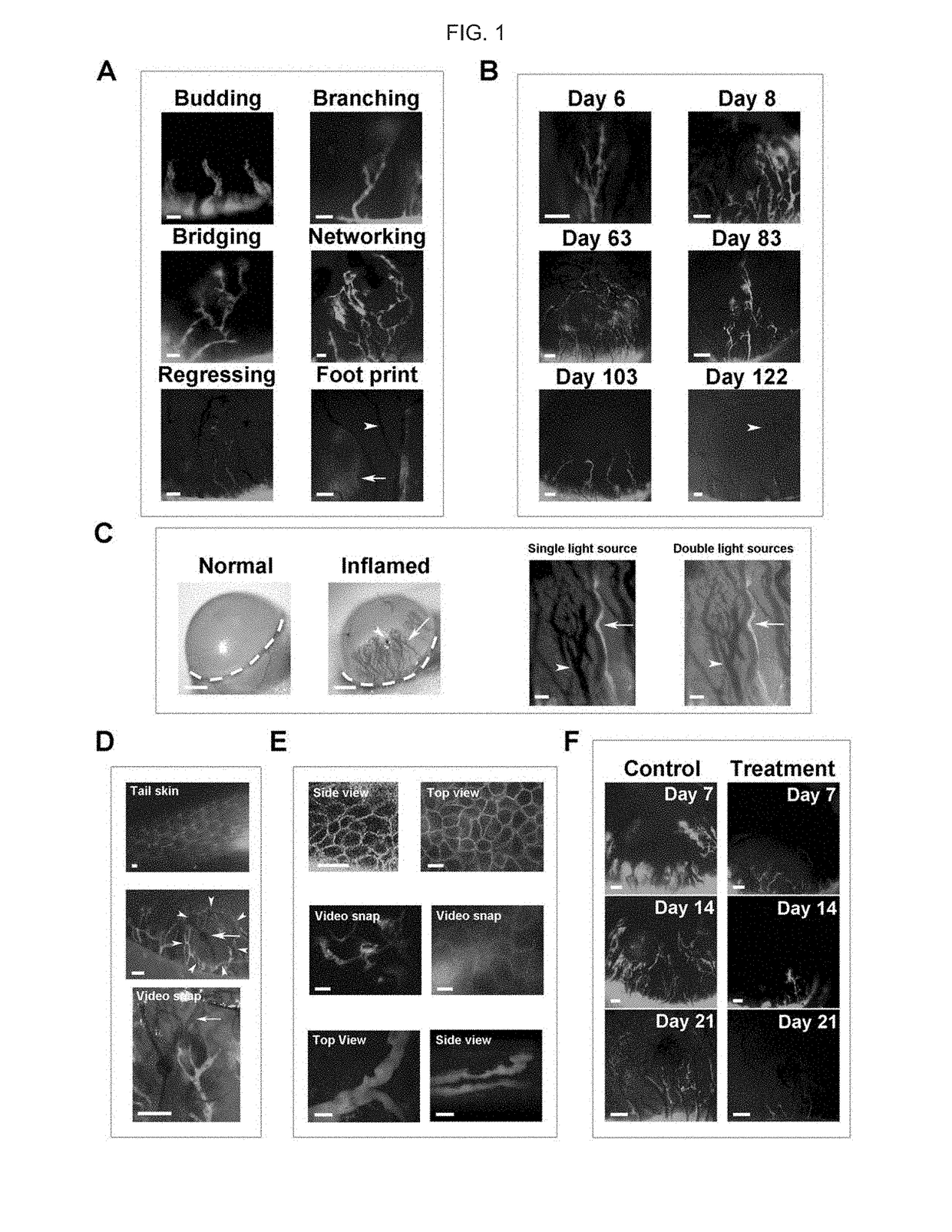
[0089]The advantages of live imaging of lymphatic vessels in the cornea over other non-transparent tissues, such as the skin, which is normally endowed with lymphatic vessels, were demonstrated. As shown in FIG. 1, Panel D, live imaging of tail skin, a site used for lymphatic imaging in previous studies, revealed a cross-sectional view of the lymphatics, which was obscured by a layer of body hairs (FIG. 1, Panel D, upper image). In contrast, imaging within the cornea, a transparent tissue that is free of any pre-existing or background vessels, showed an entire tree of newly formed lymphatic vessels from their peripheral roots to central branches (FIG. 1, Panel D, middle image). This method also enabled the observation of the lymphatic vessels in the context of their local and physiological environment, which also harbored newly formed blood vessels and the site of pa...


PUM


Abstract

Aspects of the invention include methods for visualizing lymphatic tissue in an ocular region, e.g., the cornea, of a living subject, such as a mouse or human. In certain embodiments, the methods include contacting the region with a fluorescently-labeled lymphatic tissue-specific dye, e.g., FITC or rhodamine labeled dextran, and detecting the labeled dye to visualize lymphatic tissue in the region, where the visualization may vary, from an image obtained at a single time to a video over a period of time. The invention finds use in a variety of different applications, including research and therapeutic applications.

Description

CROSS REFERENCE TO RELATED APPLICATIONS[0001]This application claims priority to the filing date of U.S. Provisional Patent Application Ser. No. 61 / 720,764, filed on Oct. 31, 2012, the disclosure of which application is herein incorporated by reference in its entirety.STATEMENT REGARDING FEDERALLY SPONSORED RESEARCH[0002]This invention was made with government support under EY017392 awarded by the National Institutes of Health. The government has certain rights in the invention.INTRODUCTION[0003]Lymphatic vessel research represents an explosive field of new discovery in recent years. Compared to blood vessels which have been studied intensively in the past, lymphatic research has suffered centuries of ignorance due to the lack of a convenient method to identify lymphatic vessels. Unlike blood vessels, lymphatic vessels are not easily visible. Accompanying the blood circulation, the lymphatic network penetrates most tissues in the body and plays critical roles in a broad spectrum of ...

Claims


Application Information

Patent Timeline
Patent Type & Authority Patents(United States)
IPC IPC(8): A61B5/00
CPCA61B5/414A61B5/0071A61B5/418A61B2503/40
Inventor CHEN, LU
Owner RGT UNIV OF CALIFORNIA
Why Eureka
Try Eureka