In vivo visualization of lymphatic tissue

a lymphatic tissue and in vivo technology, applied in the field of in vivo visualization of lymphatic tissue, can solve the problems of poor lymphatic disease treatment effect, long time-consuming and laborious, and long time-consuming, and achieve the effects of improving the quality of li

Active Publication Date: 2014-05-01
RGT UNIV OF CALIFORNIA
View PDF0 Cites 9 Cited by
  • Summary
  • Abstract
  • Description
  • Claims
  • Application Information

AI Technical Summary

Benefits of technology

[0009]In some embodiments, the polysaccharide has a molecular weight ranging from 1,000 up to 10,000 kDa. In some embodiments, the physiologically-acceptable polysaccharide is dextran. In some embodiments, visualizing the lymphatic tissue involves ob

Problems solved by technology

Compared to blood vessels which have been studied intensively in the past, lymphatic research has suffered centuries of ignorance due to the lack of a convenient method to identify lymphatic vessels.
These disorders can be disabling, disfiguring, and even life threatening.
To date, there is still little effective treatment for lymphatic disorders, so it is a field with an urgent demand for new experimental approaches and therapeutic protocols.
The burdens associated with lymphatic diseases are immense.
However, both vessel types are induced into the cornea after an inflammatory, infectious, traumatic, toxic or chemical damage event.
Despite the recent advances in corneal lymphatic research, further in-depth investigation has been greatly hampered by serious technical limitations.
For example, the conventional immunohistochemistry method which has been heavily relied upon to identify lymp

Method used


Image

  • In vivo visualization of lymphatic tissue
  • In vivo visualization of lymphatic tissue
  • In vivo visualization of lymphatic tissue

Examples


example 1

A. Example 1

Visualization of Lymphatic Vessels in the Cornea of a Subject

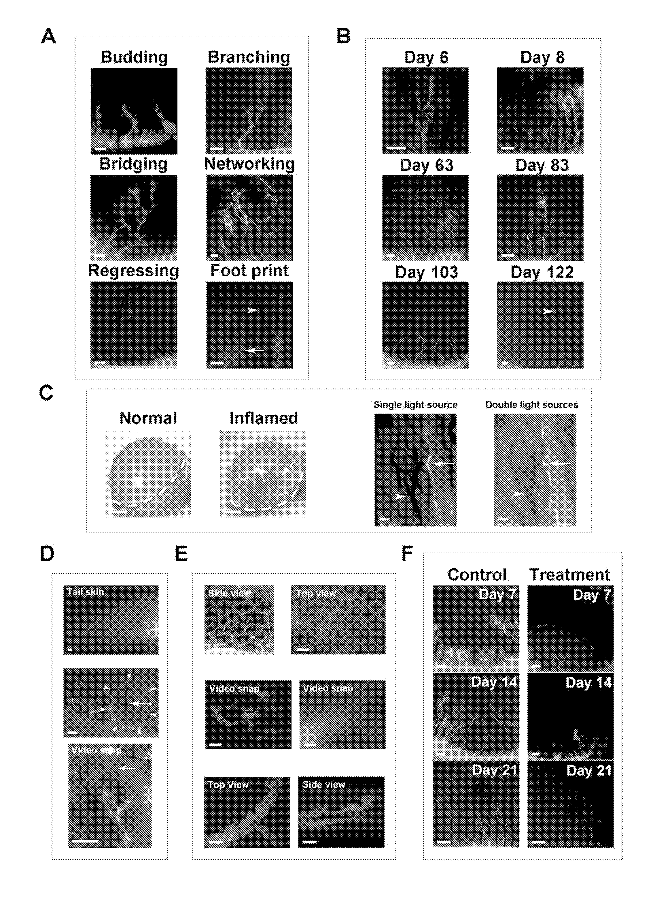
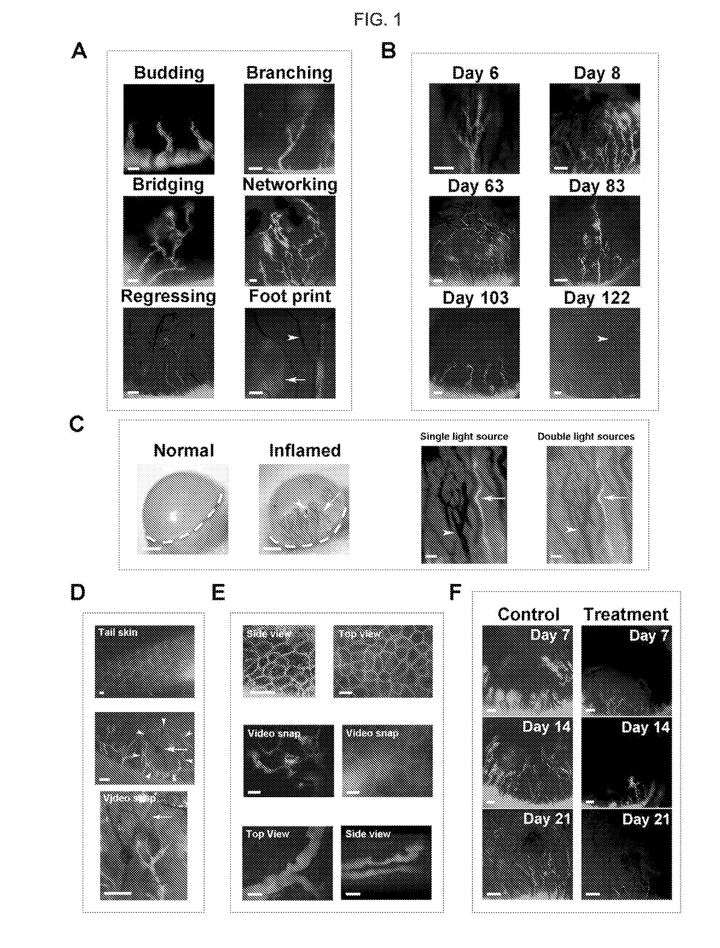
[0092]To induce inflammatory lymphangiogenesis and hemangiogenesis in the cornea, a suture protocol was employed, as described above. Corneal lymphatic vessels were visualized by subconjunctival injection of large molecular weight fluorescein isothiocyanate (FITC)-labeled dextran. The dye uptake was continuously monitored under a custom-built live imaging system with an adjustable eye and head holder to secure steady pictures while the mouse was breathing. Two types of live imaging systems were tested: 1) a non-contact live imaging system with a fluorescent stereomicroscope, and 2) a more advanced live imaging system with a two-photon excitation microscope. As demonstrated in FIG. 1, while the non-contact system was able to produce high quality images (Panels A-D, F) and a video without touching the cornea, the highly advanced two-photon system yielded more depth-sectioning and cellular details of the tissue an...

example 2

B. Example 2

Visualization of Both Lymphatic Vessels and Blood Vessels in the Cornea of a Subject

[0095]Since blood and lymphatic vessels often accompany each other, and it is important to evaluate both structures in many studies, the advantage of the subject methods over ophthalmic slit-lamp microscopy, which is commonly used in both research laboratories and clinics, was demonstrated. As demonstrated in FIG. 1, Panel C, while only blood vessels (arrow) were detectable by slit-lamp microscopy (left two images), both blood (arrowheads) and lymphatic vessels (arrows) were clearly revealed by the subject methods (right two images). While blood vessels appeared in dark shadows under the FITC fluorescence excitation source alone, they showed up in their natural red color under the combined light sources in an LED bright field view. Moreover, the images obtained by the subject methods were of better resolution and quality. The FITC-dextran labeled vessels in live corneas expressed LYVE-1, ...

example 3

C. Example 3

Advantages of Live Imaging of Lymphatic Vessels in the Cornea Over Other Non-Transparent Tissues, Such as the Skin

[0096]The advantages of live imaging of lymphatic vessels in the cornea over other non-transparent tissues, such as the skin, which is normally endowed with lymphatic vessels, were demonstrated. As shown in FIG. 1, Panel D, live imaging of tail skin, a site used for lymphatic imaging in previous studies, revealed a cross-sectional view of the lymphatics, which was obscured by a layer of body hairs (FIG. 1, Panel D, upper image). In contrast, imaging within the cornea, a transparent tissue that is free of any pre-existing or background vessels, showed an entire tree of newly formed lymphatic vessels from their peripheral roots to central branches (FIG. 1, Panel D, middle image). This method also enabled the observation of the lymphatic vessels in the context of their local and physiological environment, which also harbored newly formed blood vessels and the si...


PUM


Abstract

Aspects of the invention include methods for visualizing lymphatic tissue in an ocular region, e.g., the cornea, of a living subject, such as a mouse or human. In certain embodiments, the methods include contacting the region with a fluorescently-labeled lymphatic tissue-specific dye, e.g., FITC or rhodamine labeled dextran, and detecting the labeled dye to visualize lymphatic tissue in the region, where the visualization may vary, from an image obtained at a single time to a video over a period of time. The invention finds use in a variety of different applications, including research and therapeutic applications.

Description

CROSS REFERENCE TO RELATED APPLICATIONS[0001]This application claims priority to the filing date of U.S. Provisional Patent Application Ser. No. 61 / 720,764, filed on Oct. 31, 2012, the disclosure of which application is herein incorporated by reference in its entirety.STATEMENT REGARDING FEDERALLY SPONSORED RESEARCH[0002]This invention was made with government support under Grant No. NIH EY017392 awarded by the National Institutes of Health. The government has certain rights in the invention.INTRODUCTION[0003]Lymphatic vessel research represents an explosive field of new discovery in recent years. Compared to blood vessels which have been studied intensively in the past, lymphatic research has suffered centuries of ignorance due to the lack of a convenient method to identify lymphatic vessels. Unlike blood vessels, lymphatic vessels are not easily visible. Accompanying the blood circulation, the lymphatic network penetrates most tissues in the body and plays critical roles in a broa...

Claims


Application Information

Patent Timeline
IPC IPC(8): A61B5/00A61B19/00
CPCA61B5/0071A61B5/418A61B19/54A61B5/414A61B2503/40
Inventor CHEN, LUYUEN, DON
Owner RGT UNIV OF CALIFORNIA
Why Eureka
Try Eureka