Method and system for segmentation of brain structures in 3D magnetic resonance images

a brain structure and 3d magnetic resonance imaging technology, applied in the field of medical imaging of the brain, can solve the problems of inability to use conventional techniques for segmenting sub-cortical and cortical brain structures for brain growth, abnormal brain pattern detection, and brain growth

Active Publication Date: 2017-10-31
SIEMENS HEALTHCARE GMBH
View PDF11 Cites 1 Cited by
  • Summary
  • Abstract
  • Description
  • Claims
  • Application Information

AI Technical Summary

Problems solved by technology

Detection and delineation of sub-cortical and cortical brain structures in magnetic resonance (MR) data is an important problem in medical imaging analysis.
Manual delineation of such sub-cortical and cortical brain structures in 3D MR data is a challenging task, ever for expert users, such as clinicians or neuroanatomists.
Moreover, even for experienced users, the manual delineation of a single brain structure is a time-consuming process, and manual annotations may vary significantly among experts as a result of individual experience and interpretation.
Conventional techniques for segmenting sub-cortical and cortical brain structures cannot be used for purposes such as detecting abnormal brain patterns, studying brain diseases, and studying brain growth due to a lack of proper generalization.
In particular, conventional methods may successfully perform segmentation of sub-cortical and cortical brain structures in MR data for healthy adult brains, but cannot reliably detect abnormal brain structures or brain structures in pediatric brains.

Method used


Image

  • Method and system for segmentation of brain structures in 3D magnetic resonance images
  • Method and system for segmentation of brain structures in 3D magnetic resonance images
  • Method and system for segmentation of brain structures in 3D magnetic resonance images

Examples


Embodiment Construction

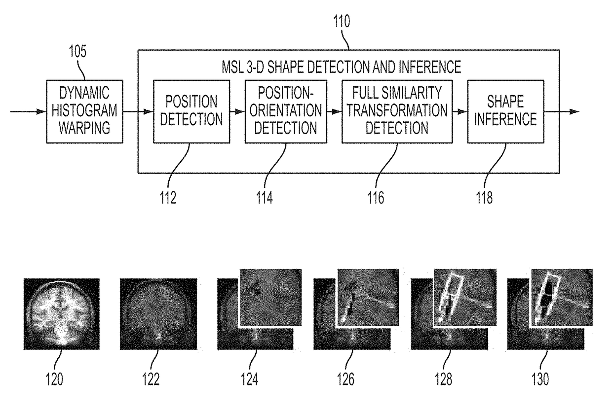
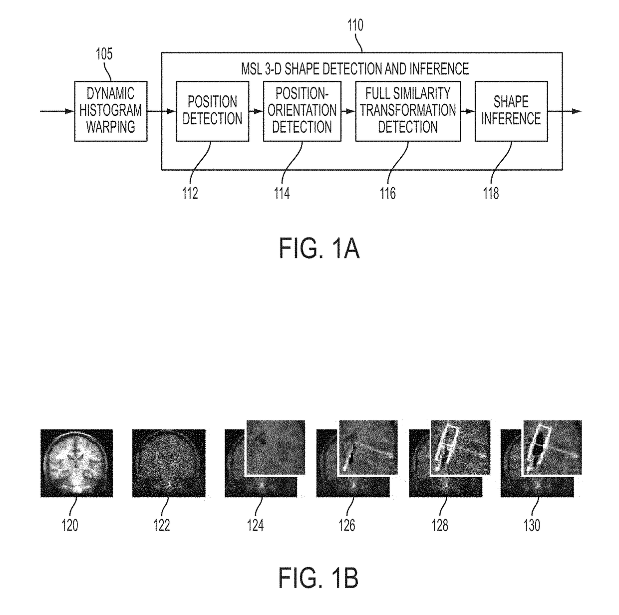
[0014]The present invention is directed to a method and system for automatic segmentation of sub-cortical and cortical brain structures in 3D magnetic resonance (MR) images. Embodiments of the present invention are described herein to give a visual understanding of the sub-cortical and cortical brain structure segmentation method. A digital image is often composed of digital representations of one or more objects (or shapes). The digital representation of an object is often described herein in terms of identifying and manipulating the objects. Such manipulations are virtual manipulations accomplished in the memory or other circuitry / hardware of a computer system. Accordingly, is to be understood that embodiments of the present invention may be performed within a computer system using data stored within the computer system.

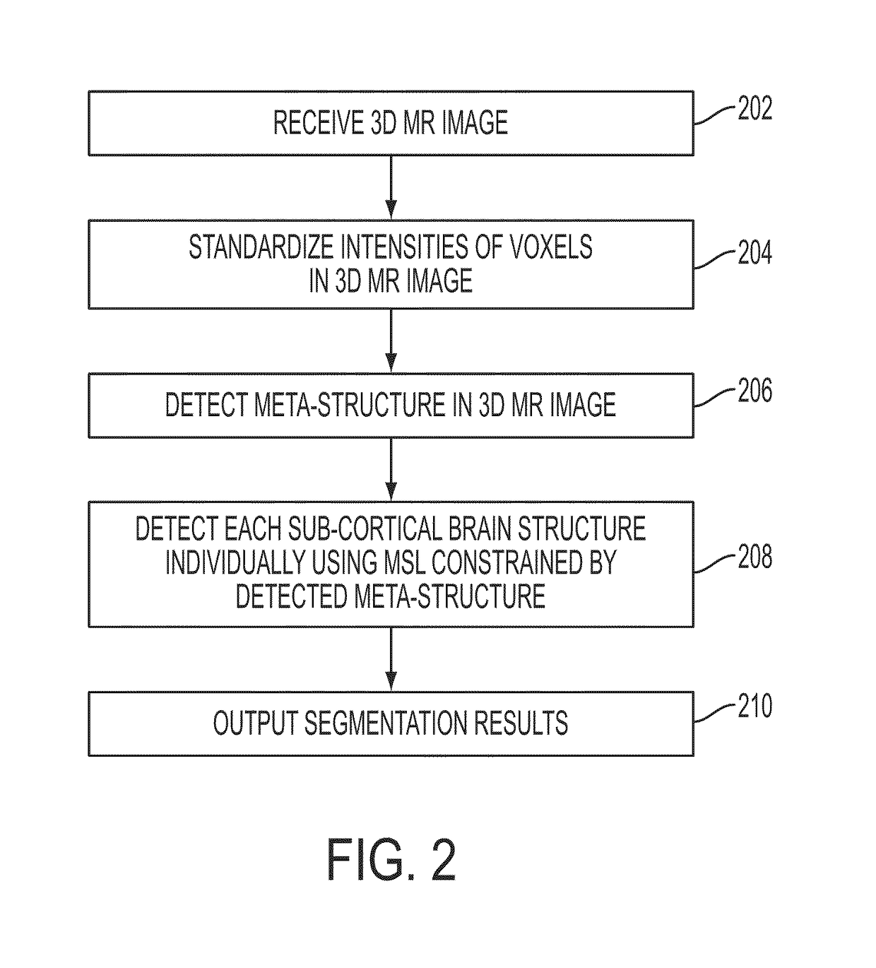
[0015]Embodiments of the present invention are directed to automated (sub)-cortical brain structure segmentation in 3D MR images. As used herein, a “(sub)-cortical...


PUM


Abstract

A method and system for segmenting multiple brain structures in 3D magnetic resonance (MR) images is disclosed. After intensity standardization of a 3D MR image, a meta-structure including center positions of multiple brain structures is detected in the 3D MR image. The brain structures are then individually segmented using marginal space learning (MSL) constrained by the detected meta-structure.

Description

[0001]This application claims the benefit of U.S. Provisional Application No. 61 / 098,273, filed Sep. 19, 2008, the disclosure of which is herein incorporated by reference.BACKGROUND OF THE INVENTION[0002]The present invention relates to medical imaging of the brain, and more particularly, to automatic segmentation of sub-cortical and cortical brain structures in 3D magnetic resonance images.[0003]Detection and delineation of sub-cortical and cortical brain structures in magnetic resonance (MR) data is an important problem in medical imaging analysis. For example, delineated sub-cortical and cortical brain structures can be used in detecting abnormal brain patterns, studying various brain diseases, and studying brain growth. Manual delineation of such sub-cortical and cortical brain structures in 3D MR data is a challenging task, ever for expert users, such as clinicians or neuroanatomists. Task-specific protocols for manual brain structure annotation exist, but the correct utilizati...

Claims


Application Information

Patent Timeline
Patent Type & Authority Patents(United States)
IPC IPC(8): G06T7/143G06T7/11
CPCG06T7/143G06T7/11G06T2207/30016G06T2207/10088
Inventor WELS, MICHAELCARNEIRO, GUSTAVO HENRIQUE MONTEIRO DE BARROSHUBER, MARTINCOMANICIU, DORINZHENG, YEFENG
Owner SIEMENS HEALTHCARE GMBH
Why Eureka
Try Eureka