Method and system for automatic prostate segmentation in magnetic resonance images

a magnetic resonance image and automatic segmentation technology, applied in the field of medical imaging of the prostate, can solve the problems of computational cost and difficult automatic segmentation of the prostate in mr images, and achieve the effect of short processing time and efficient radiation therapy planning

Inactive Publication Date: 2016-02-23
SIEMENS HEALTHCARE GMBH
View PDF11 Cites 1 Cited by
  • Summary
  • Abstract
  • Description
  • Claims
  • Application Information

AI Technical Summary

Benefits of technology

[0005]The present invention provides a method and system for automatically segmenting the prostate in magnetic resonance (MR) images. Embodiments of the present invention provide a fully automatic segmentation of the prostate in an MR image with a relatively short processing time so that the segmentation results can be efficiently used for radiation therapy planning. Instead of using an explicit registration of images, embodiments of the present invention compactly represent a training set of images with discriminative classifiers that are used to align a statistical mesh model of the prostate onto an MR image. Rather than relying on edge detectors or hand-designed features, embodiments of the present invention utilize classifiers that aggregate and choose the best image features from a large feature pool.

Problems solved by technology

Detection and delineation of the prostate in medical image data is an important problem in medical imaging analysis.
However, automated segmentation of the prostate in MR images is challenging due to global inter-scan variability and intra-scan intensity variation caused by an endorectal coil, as well as due to the similar appearance of the prostate and the seminal vesicles at the apex, geometric variability due to disease, and adjacent structures such as the rectum and the bladder.
Although such registrations methods perform well when one of the templates is similar to the target image, they are often computationally expensive, typically requiring several minutes to return a segmentation.

Method used


Image

  • Method and system for automatic prostate segmentation in magnetic resonance images
  • Method and system for automatic prostate segmentation in magnetic resonance images
  • Method and system for automatic prostate segmentation in magnetic resonance images

Examples


Embodiment Construction

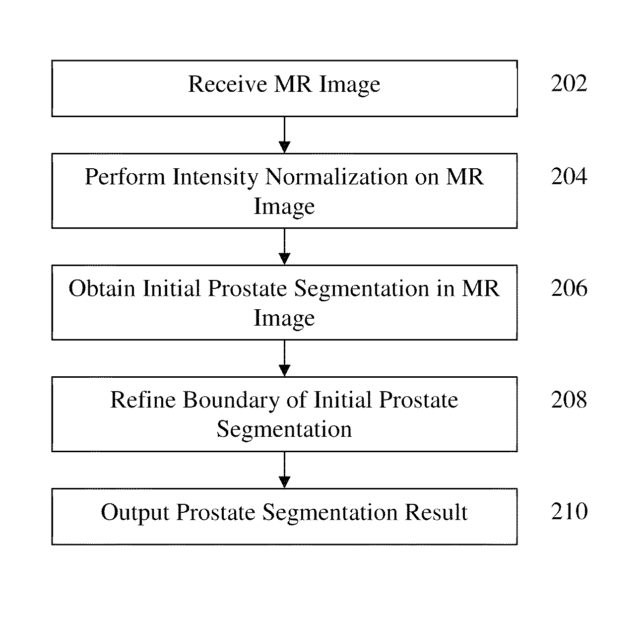
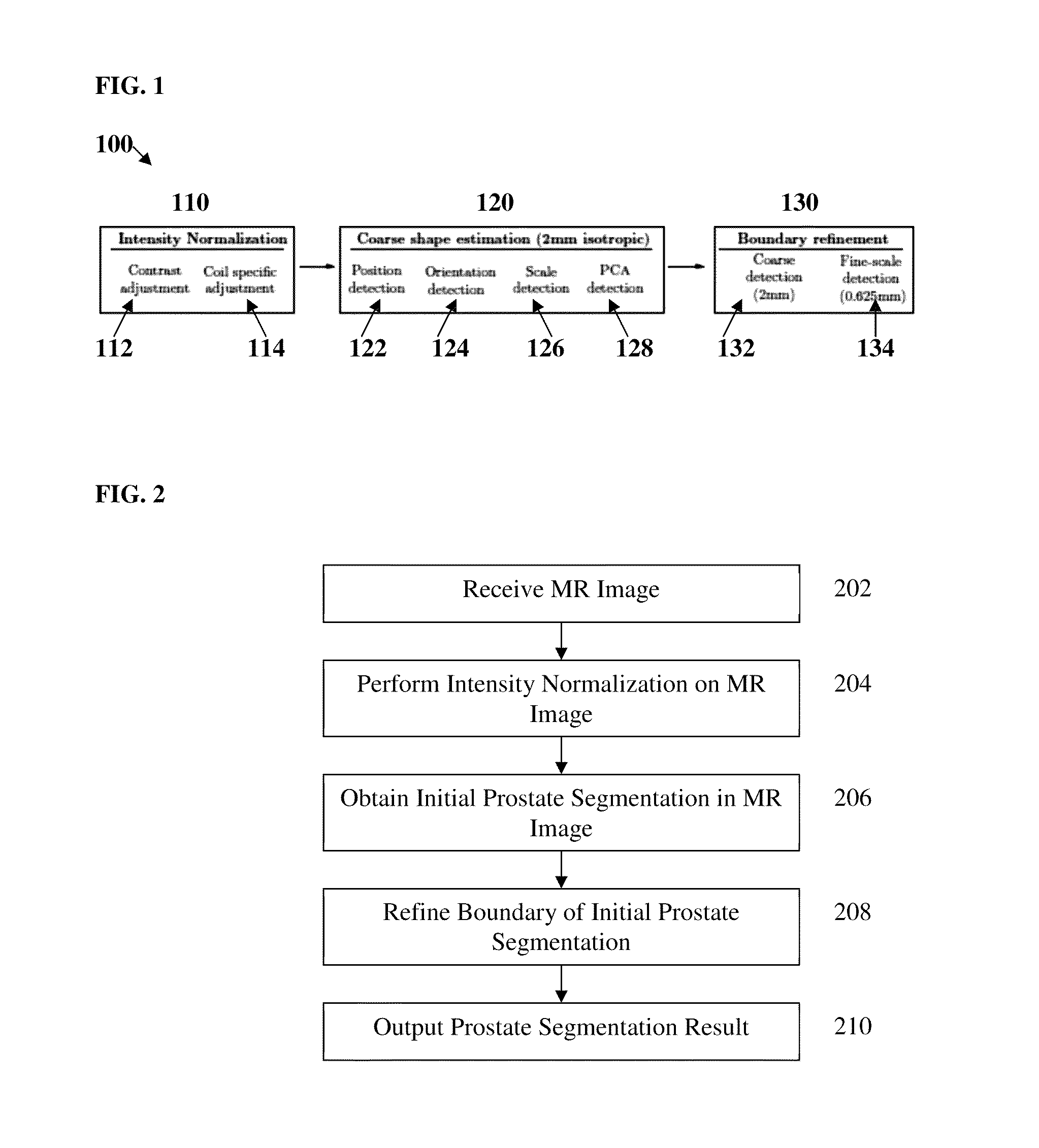
[0015]The present invention is directed to a method and system for fully automatic segmentation of the prostate in magnetic resonance (MR) images. Embodiments of the present invention are described herein to give a visual understanding of the prostate segmentation method. A digital image is often composed of digital representations of one or more objects (or shapes). The digital representation of an object is often described herein in terms of identifying and manipulating the objects. Such manipulations are virtual manipulations accomplished in the memory or other circuitry / hardware of a computer system. Accordingly, is to be understood that embodiments of the present invention may be performed within a computer system using data stored within the computer system.

[0016]Embodiments of the present invention are directed to automated prostate segmentation in MR images. Embodiments of the present invention utilize learning-based methods and hierarchical boundary definition for efficient...


PUM


Abstract

A method and system for fully automatic segmentation the prostate in magnetic resonance (MR) image data is disclosed. Intensity normalization is performed on an MR image of a patient to adjust for global contrast changes between the MR image and other MR scans and to adjust for intensity variation within the MR image due to an endorectal coil used to acquire the MR image. An initial prostate segmentation in the MR image is obtained by aligning a learned statistical shape model of the prostate to the MR image using marginal space learning (MSL). The initial prostate segmentation is refined using one or more trained boundary classifiers.

Description

[0001]This application claims the benefit of U.S. Provisional Application No. 61 / 674,912, filed Jul. 24, 2012, the disclosure of which is herein incorporated by reference.BACKGROUND OF THE INVENTION[0002]The present invention relates to medical imaging of the prostate, and more particularly, to automatic segmentation of the prostate in magnetic resonance images.[0003]Detection and delineation of the prostate in medical image data is an important problem in medical imaging analysis. For example, prostate cancer is often treated by radiation therapy where precise targeting of the prostate is important. The improved contrast of magnetic resonance (MR) abdominal image over other imaging modalities affords more accurate and consistent manual contouring of the prostate. Consequently, MR imaging now plays an increasing role in prostate radiotherapy planning. Accordingly, automated methods for segmenting abdominal structures in MR are desirable to improve the clinical workflow. However, aut...

Claims


Application Information

Patent Timeline
Patent Type & Authority Patents(United States)
IPC IPC(8): G06T7/00G06T5/00
CPCG06T7/0081G06T5/009G06T7/0089G06T2200/04G06T2207/10088G06T2207/20016G06T2207/20081G06T2207/30081G06T7/11G06T7/149
Inventor BIRKBECK, NEILZHANG, JINGDANREQUARDT, MARTINKIEFER, BERTHOLDGALL, PETERZHOU, SHAOHUA KEVIN
Owner SIEMENS HEALTHCARE GMBH
Why Eureka
Try Eureka