Method and system for segmentation of the prostate in 3D magnetic resonance images

a magnetic resonance image and prostate technology, applied in the field of medical imaging of the prostate, can solve the problems of significant inter- and intra-user variability, time-consuming and labor-intensive manual delineation of the prostate, and difficult and labor-intensive task, and achieve the effect of short processing tim

Active Publication Date: 2015-05-05
SIEMENS HEALTHCARE GMBH
View PDF20 Cites 2 Cited by
  • Summary
  • Abstract
  • Description
  • Claims
  • Application Information

AI Technical Summary

Benefits of technology

[0005]The present invention provides a method and system for automatically segmenting the prostate in multi-spectral 3D MR images. Embodiments of the present invention provide a fully automatic segmentation of the prostate with a relatively short processing time that works with several types of data including pathological cases, and provides a segmentation accuracy that allows the segmentation results to be suitable for inverse radiation therapy planning.

Problems solved by technology

Detection and delineation of the prostate in magnetic resonance (MR) and computed tomography (CT) images is an important problem in medical imaging analysis.
Manual delineation of the prostate in 3D MR data, sometimes in combination with 3D CT data, is a challenging and labor intensive task, even for expert radiologists.
The manual delineation of the prostate is a time-consuming process, and there is significant inter- and intra-user variability among manual segmentations of the prostate.

Method used


Image

  • Method and system for segmentation of the prostate in 3D magnetic resonance images
  • Method and system for segmentation of the prostate in 3D magnetic resonance images
  • Method and system for segmentation of the prostate in 3D magnetic resonance images

Examples


Embodiment Construction

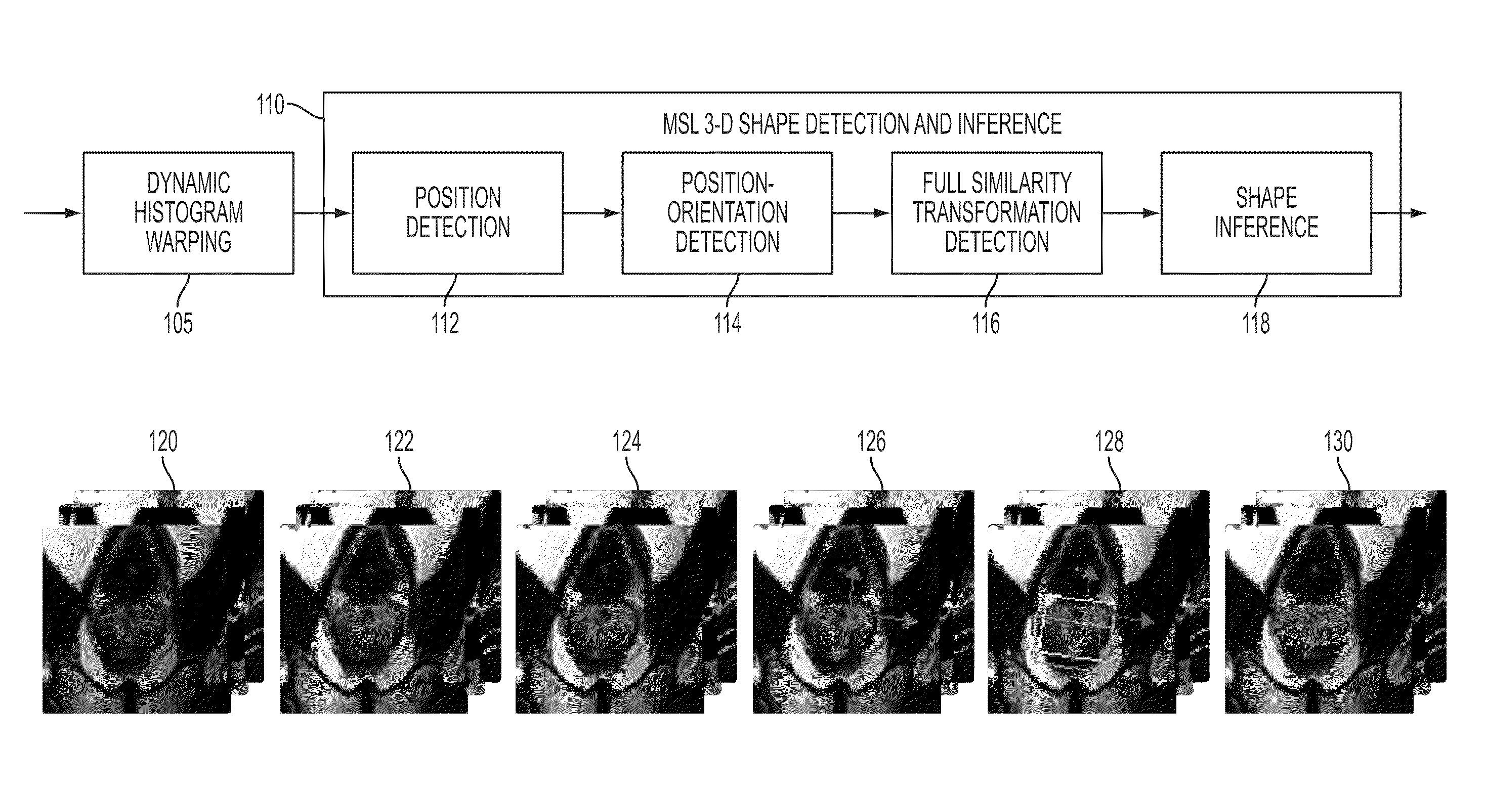
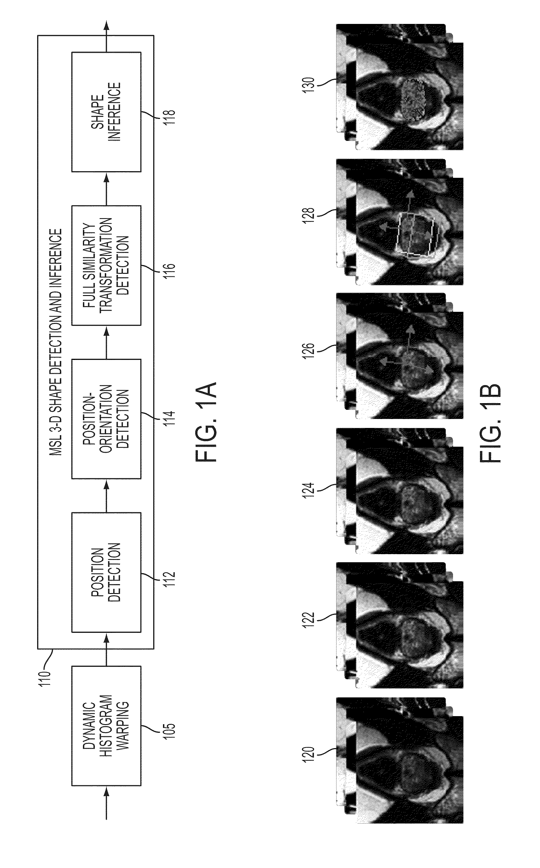
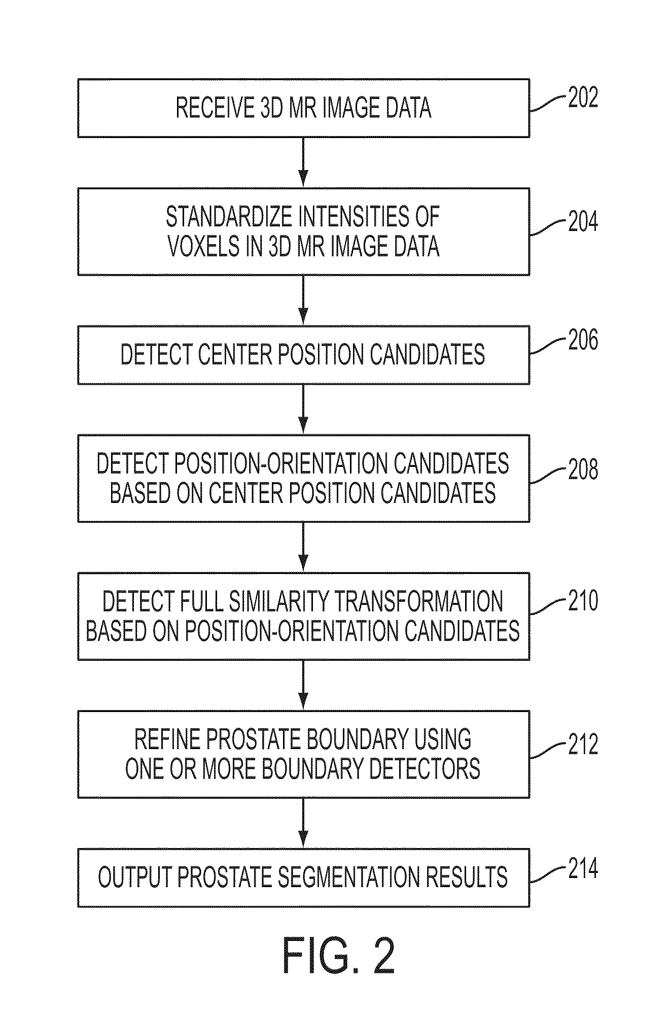
[0014]The present invention is directed to a method and system for fully automatic segmentation of the prostate in multi-spectral 3D magnetic resonance (MR) images. A multi-spectral MR image is composed of vector-valued intensity values, i.e., one or more scalar intensity values per voxel. Different MR channels (T1-weighted, T2-weighted, etc.) may emphasize on different tissue characteristics providing additional information about the depicted image content. Embodiments of the present invention are described herein to give a visual understanding of the prostate segmentation method. A digital image is often composed of digital representations of one or more objects (or shapes). The digital representation of an object is often described herein in terms of identifying and manipulating the objects. Such manipulations are virtual manipulations accomplished in the memory or other circuitry / hardware of a computer system. Accordingly, is to be understood that embodiments of the present inve...


PUM


Abstract

A method and system for fully automatic segmentation the prostate in multi-spectral 3D magnetic resonance (MR) image data having one or more scalar intensity values per voxel is disclosed. After intensity standardization of multi-spectral 3D MR image data, a prostate boundary is detected in the multi-spectral 3D MR image data using marginal space learning (MSL). The detected prostate boundary is refined using one or more trained boundary detectors. The detected prostate boundary can be split into patches corresponding to anatomical regions of the prostate and the detected prostate boundary can be refined using trained boundary detectors corresponding to the patches.

Description

[0001]This application claims the benefit of U.S. Provisional Application No. 61 / 262,330, filed Nov. 18, 2009, the disclosure of which is herein incorporated by reference.BACKGROUND OF THE INVENTION[0002]The present invention relates to medical imaging of the prostate, and more particularly, to fully automatic segmentation of the prostate in 3D magnetic resonance images.[0003]Detection and delineation of the prostate in magnetic resonance (MR) and computed tomography (CT) images is an important problem in medical imaging analysis. For example, prostate cancer is often treated by radiation therapy where precise targeting of the prostate is important. The exposure of neighboring organs, such as the bladder or the rectum, to ionizing radiation should be reduced to an absolute minimum. For this purpose, accurate localization of the prostate by exact organ delineation is critical. In addition, prostate delineation in medical images can be used for MR spectroscopy analysis (MRSI) to obtai...

Claims


Application Information

Patent Timeline
Patent Type & Authority Patents(United States)
IPC IPC(8): G06K9/00G06K9/62G06T7/00
CPCG06K9/6207G06T7/0012G06T7/0083G06T7/0089G06T2207/10081G06T2207/10088G06T2207/30081G06T7/12G06T7/149G06V10/755
Inventor WELS, MICHAELSUEHLING, MICHAELKELM, MICHAELSEIFERT, SASCHACOSTA, MARIA JIMENACAVALLARO, ALEXANDERHUBER, MARTINCOMANICIU, DORIN
Owner SIEMENS HEALTHCARE GMBH
Why Eureka
Try Eureka