
Platelet analysis

a platelet and analysis method technology, applied in the field of platelet analysis, can solve the problems of difficult to accurately visually monitor platelets, difficult to accurately analyse a large number of platelets using such known platelet adhesion assays, and no adhesion assay available to analyse, so as to prevent further platelet activation, reduce the difficulty of detection, and improve the effect of detection efficiency

Inactive Publication Date: 2018-04-10
DUBLIN CITY UNIVERSITY +1
View PDF7 Cites 0 Cited by
  • Summary
  • Abstract
  • Description
  • Claims
  • Application Information

AI Technical Summary

Benefits of technology

[0011]Any fluid composition that contains platelets may be analysed using the above method. Thus, extracts of whole blood that contain platelets (including suspensions of platelets in a physiological fluid carrier), diluted blood, or the like, may be used. A particular advantage of the present invention is the fact that, unlike prior art methods where extracts or dilutions of whole blood must be used, the present method may utilise whole blood (which does not require dilution).
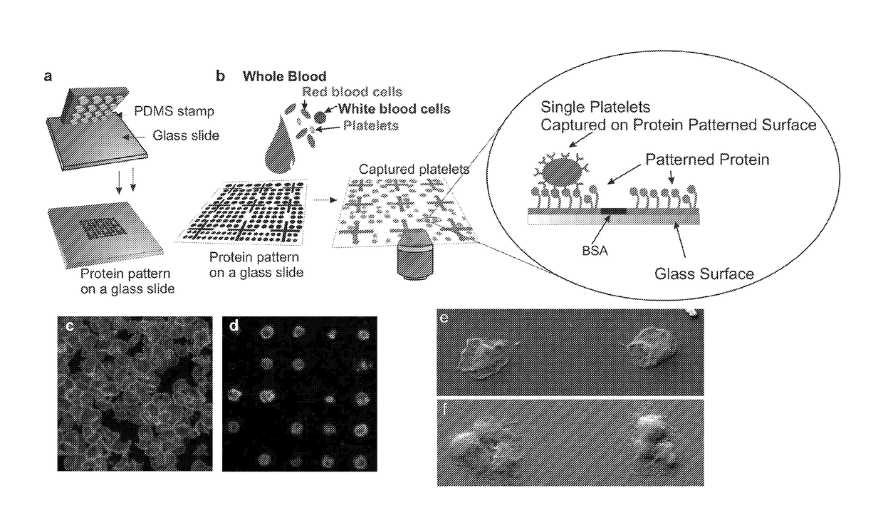
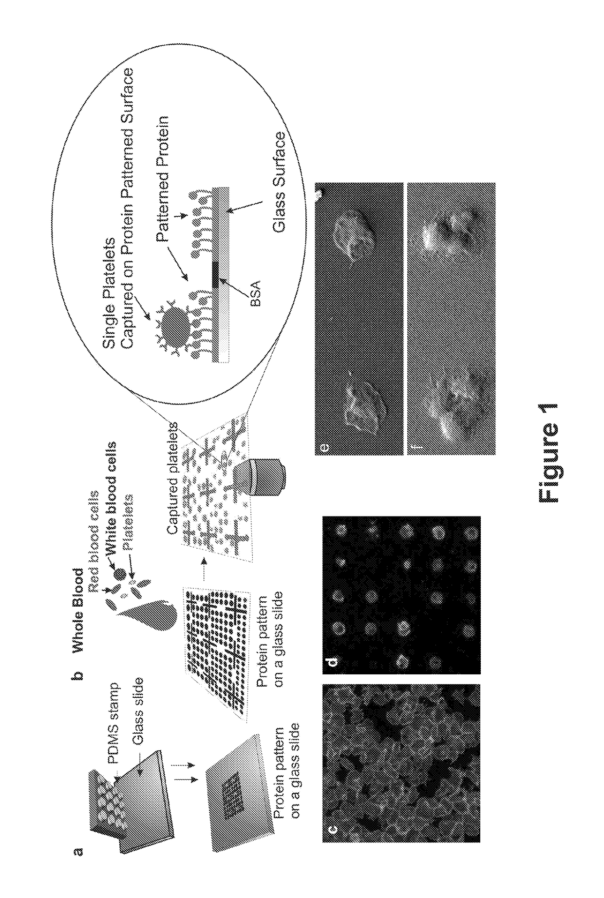
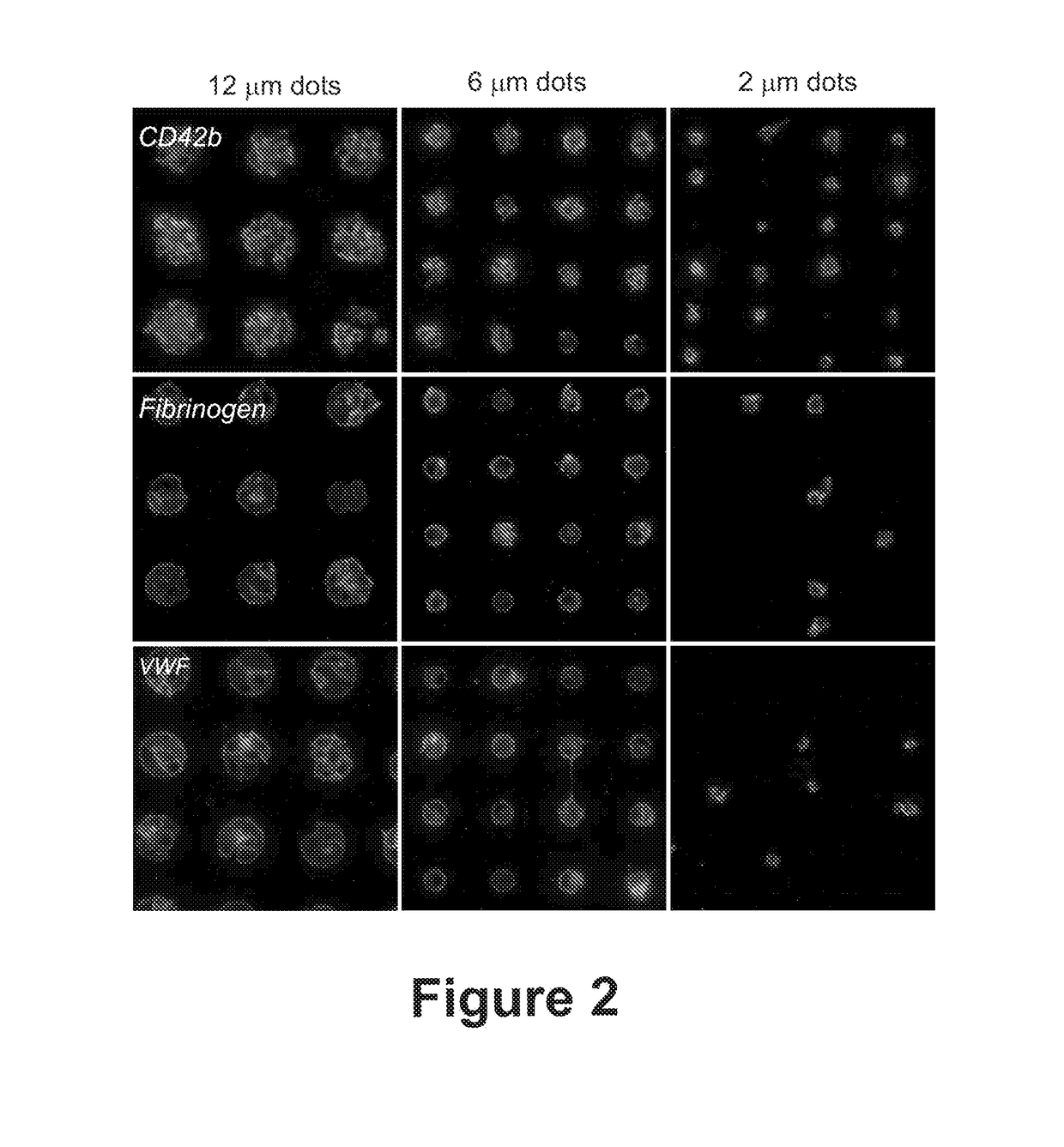
[0012]Additives such as anticoagulant additives (e.g. citrate) may be added to the fluid composition. When the method is used for diagnostic purposes, or when the efficacy of a therapeutic is being tested, additives such as anticoagulants, platelet binding agents such as anti-platelet antibodies or platelet antagonists, or the therapeutic to be tested, may be added to the fluid composition.
[0013]An additional advantage of the method according to the present invention is the fact that it can be operated using relatively small amounts of fluid composition. A sample of fluid composition of less than 2, 1, 0.5. 0.25, 0.1, or 0.01 ml may be used in the method. Preferably the volume of fluid composition used in the method is from 1 μl to 1 ml.
[0014]In order for platelets to bind preferentially to the platelet-binding zones, an embodiment of the present invention includes platelet-binding zones that comprise platelet-affinity binding agents that are bound to the surface of the substrate. Preferably, the platelet-affinity binding agents are specific for platelet binding, and so may be specific for membrane-bound platelet-specific proteins. The person skilled in the art would be well aware of the many binding partners that would be suitable for binding platelets. Not wishing to be restricted further, but in the interests of clarity, the platelet-affinity binding agents may comprise fibrinogen, von Willebrand factor, collagen, platelet-specific antibodies, proteins, peptides, aptamers or any fragments thereof capable of binding platelets, or the like, or any combination thereof. Binding between platelet and platelet-affinity binding agent is preferably strong enough that a majority of the bound platelets remain bound for at least about 1 minute, (though shorter binding times could be supported in special circumstances, especially where desorption rate may be of interest—e.g., platelets on vWF under shear). Thus, according to one embodiment of the present invention, prior to step a. the platelet-affinity binding agents are applied to the substrate. Any method of application that enables the platelet-affinity binding agents to be applied to the substrate as a regular distribution of platelets according to the aforementioned embodiments are preferred, for example:—by microcontact printing, light-directed spatially addressable parallel chemical synthesis, photo-patterning, nano dip-pen patterning, piezo-droplet technique or any combination thereof.
[0015]Optionally, visualisation agents are applied to the substrate together with the platelet-affinity binding agents. Visualisation agents may be any agent, such as radioactive markers or, fluorescent or coloured dyes, that can readily be visualised (i.e. detected for analysis, by simply being viewed by naked eye or via a microscope, or automated detection where an emission such as light, radioactivity etc. is detected by a machine). The visualisation agents may selectively bind the platelet-affinity binding agents, and may comprise, for example a platelet-affinity binding agent antibody bound to a radioactive markers or, fluorescent or coloured dye. This enables the pattern of deposits of platelet-affinity binding agents (and so position, shape and size of platelet binding zones) to be visualised. Visualisation agents that selectively bind to the platelet-affinity binding agents may alternatively or additionally be applied to the substrate after the platelet-affinity binding agents are applied.
[0016]In order for the platelets in the fluid composition to bind to the platelet-binding zones on the substrate, the fluid composition must come into contact with the substrate. This may be achieved by simply applying the fluid composition directly to the substrate (e.g. by spotting with a pipette). Alternatively, the fluid composition may be passed over the substrate such that platelets of the composition are in contact with and bind to the platelet-binding zones. This may be achieved by creating a flow of fluid composition over the surface of the substrate; the flow being created by a pressure pushing the fluid composition over the substrate (i.e. by pumping) or by a negative pressure that pulls the composition over the substrate (i.e. by suction). The fluid composition should be in contact with the substrate for a sufficient length of time to enable binding of the platelets to the platelet-binding zones. Preferably, therefore, the fluid composition contacts the substrate for more than 10 seconds, 1 minute, 5 minutes or 10 minutes; but preferably not more than 20, 25 or 30 minutes. Contact may be terminated by applying a wash to the substrate, or by discontinuing the supply of fluid composition to the substrate. The wash should be capable of removing unbound platelets and other components of the fluid composition, but not capable of removing bound platelets. It is also preferred that the wash is an aqueous solution that is capable of preventing further platelet activation. By way of example only, a suitable wash may comprise 130 mM NaCl, 6 mM dextrose, 9 mM NaHCO3, 10 mM Na citrate, 10 mM Tris, 3 mM KCl, 0.81 mM KH2PO4, 0.9 mM MgCl2. Alternatively, or in addition, a further wash may be applied in order to fix the bound platelets (e.g. a formaldehyde or paraformaldehyde wash). Thus, it is preferred that prior to step b. the fluid composition including non-bound platelets is washed from the substrate and / or a fixing wash is applied to the bound platelets.

Problems solved by technology

However, most of these techniques, such as aggregometry or protein gels, analyse bulk samples in which a large number of platelets are present, giving as results the mean response of all the cells and not the response of individual cells.
However, this technique is only able to measure platelets remaining as single cells in solution, a condition where platelets would not be allowed to be activated in vivo and also where it is difficult to accurately visually monitor platelets.
Numerous studies have been carried out using platelet adhesion assays, but they involve the use of relatively large volumes of blood and variable platelet preparation steps which are time consuming and might induce platelet activation.
The applicants have found that it is difficult to accurately analyse a large number of platelets using such known platelet adhesion assays; only the results obtained from manual counting in a few selected areas are usually achieved, and the way of choosing the areas for analysis is commonly not clearly stated or done in a reproducible and objective way.
Nowadays there is no adhesion assay available that could analyse in an automatic way a large number of single platelets, like in flow cytometry.

Method used


Image

  • Platelet analysis
  • Platelet analysis
  • Platelet analysis

Examples


example 2

[0113]Fabrication of Single Platelet Microarrays from Whole Blood

[0114]Materials

[0115]For the fabrication of Polydimethylsiloxane (PDMS) stamps for microcontact printing, Dow Corning Sylgard 184 was purchased from Farnell (Farnell Ireland, Dublin, Ireland) MICROPOSIT™ S1818™ Positive Photoresist (Chestech Ltd, Warwickshire, UK) was used in the fabrication of masters for PDMS curing. Cover glass slips (20×20 mm) were purchased from Agar scientific ltd (Essex, England). PBS tablets, paraformaldehyde, sodium citrate and all the chemicals for the buffer were purchased from Sigma Chemical Company (St. Louis, Mo., USA). For each day, a 37% stock solution of paraformaldehyde (PFA) was made in distilled water with 1.4% 2N NaOH. A working solution of 3.7% PFA in PBS was then prepared from this. The platelet buffer A (130 mM NaCl, 6 mM dextrose, 9 mM NaHCO3, 10 mM Na citrate, 10 mM Tris, 3 mM KCl, 0.81 mM KH2PO4, 0.9 mM MgCl2) was made up from stock solutions and the pH adjusted to 7.35 on th...

example 3

[0127]Monitoring the Effect of Abciximab (Reopro®)

[0128]The single platelet counting method enables high resolution and reproducible platelet adhesion and function assays in contrast with fluorescence measurements of platelet adhesion. The drug abciximab inhibits platelet adhesion to fibrinogen. In this example (results showin in FIG. 7) it is demonstrated that the single platelet counting method enables the detection of the effect of different doses of abxicimab in the blood with high precision and reproducibility by quantification of the percentage of dots occupied in the 6 microns diameter fibrinogen dots array. We compare this platelet adhesion quantification method with a classical method which consists on the measure of the fluorescence intensity originated by the platelets adhering to the surface (which have been fluorescently labelled after adhesion took place). Blood samples pre-incubated with 1.5, 2 or 2.5 μg / mL abciximab (drug which inhibits the interaction between platel...

example 4

[0129]Platelets Adhering to Dots of Different Shapes

[0130]Four circular platelet adhesion zones of varying sizes, four square platelet adhesion zones of varying sizes, and four tear-drop shaped platelet adhesion zones of varying sizes were prepared using fibrinogen according the methods described in previous examples. Each of the platelet adhesion zones were incubated with whole blood from the same donor and the platelet binding analysed according to the previous examples. The number of platelets bound to each platelet binding zone was seen to be dictated by the size of the platelet binding zone, whilst no difference was attributed to the different shapes. See FIG. 8. This also shows that platelets captured by the adhesion zones do not spread beyond those adhesion zones, as demonstrated by the fact that the shape of the adhesion zone is followed by the bound platelets.


PUM

PropertyMeasurementUnit
surface areaaaaaaaaaaa
diameteraaaaaaaaaa

Abstract

The present invention relates to a method, including a diagnostic method, for characterising platelets. More particularly, the invention relates to methods for characterising platelets by immobilising platelets on a substrate for detection and subsequent characterisation, and to devices on which such a method may be practiced. The method comprises the steps of:—a. contacting a substrate that includes a plurality of discrete platelet-binding zones having a surface area of 7850 μm2 orless with a fluid composition comprising platelets; and b. detecting platelets bound to the platelet-binding zones and thereby characterising platelets.

Description

FIELD OF THE INVENTION[0001]The present invention relates to a method for characterising platelets. More particularly, the invention relates to methods for characterising platelets by immobilising platelets on a substrate for detection and subsequent characterisation, and to devices on which such a method may be practiced.BACKGROUND OF THE INVENTION[0002]Circulating platelets play a critical role in normal homeostasis; they adhere and aggregate at sites of vascular injury to initiate thrombus formation. Platelets also play a central role in the pathogenesis of arterial thrombosis, which accounts for the clinical events associated with cardiovascular disease. They are involved in the haematogenous spread of cancerous cells during the metastatic cascade, and in inflammatory diseases. The study of the pathways leading to platelet activation and expression of adhesive ligands is very important to understand and regulate platelet interaction with other platelets, tumour cells or leukocyt...

Claims


Application Information

Patent Timeline
Patent Type & Authority Patents(United States)
IPC IPC(8): G01N33/50
CPCG01N33/5091G01N33/5094
Inventor BASABE-DESMONTS, LOURDESRAMSTROM, SOFIARICCO, ANTONIODERMOT, KENNYMEADE, GERARDENEO'NEILL, SARAHRIAZ, ASIFLEE, LUKE
Owner DUBLIN CITY UNIVERSITY
Learn More