
Device for positioning a body part and body fluid analyzer

A body part, analyzer technology, applied in the field of analyzers

Inactive Publication Date: 2011-11-09
F HOFFMANN LA ROCHE & CO AG
View PDF4 Cites 0 Cited by
  • Summary
  • Abstract
  • Description
  • Claims
  • Application Information

AI Technical Summary

Problems solved by technology

Point-like interventions are thus required for blood collection, where there is a risk of excess blood contaminating the device
Furthermore, body parts can slip unintentionally if the coefficient of friction between the compression unit and the skin drops below a lower limit, for example due to sweating or fat deposits on the skin
If the compression unit is completely folded inside the instrument, that would also fail in this case

Method used


Image

  • Device for positioning a body part and body fluid analyzer
  • Device for positioning a body part and body fluid analyzer
  • Device for positioning a body part and body fluid analyzer

Examples


Embodiment Construction

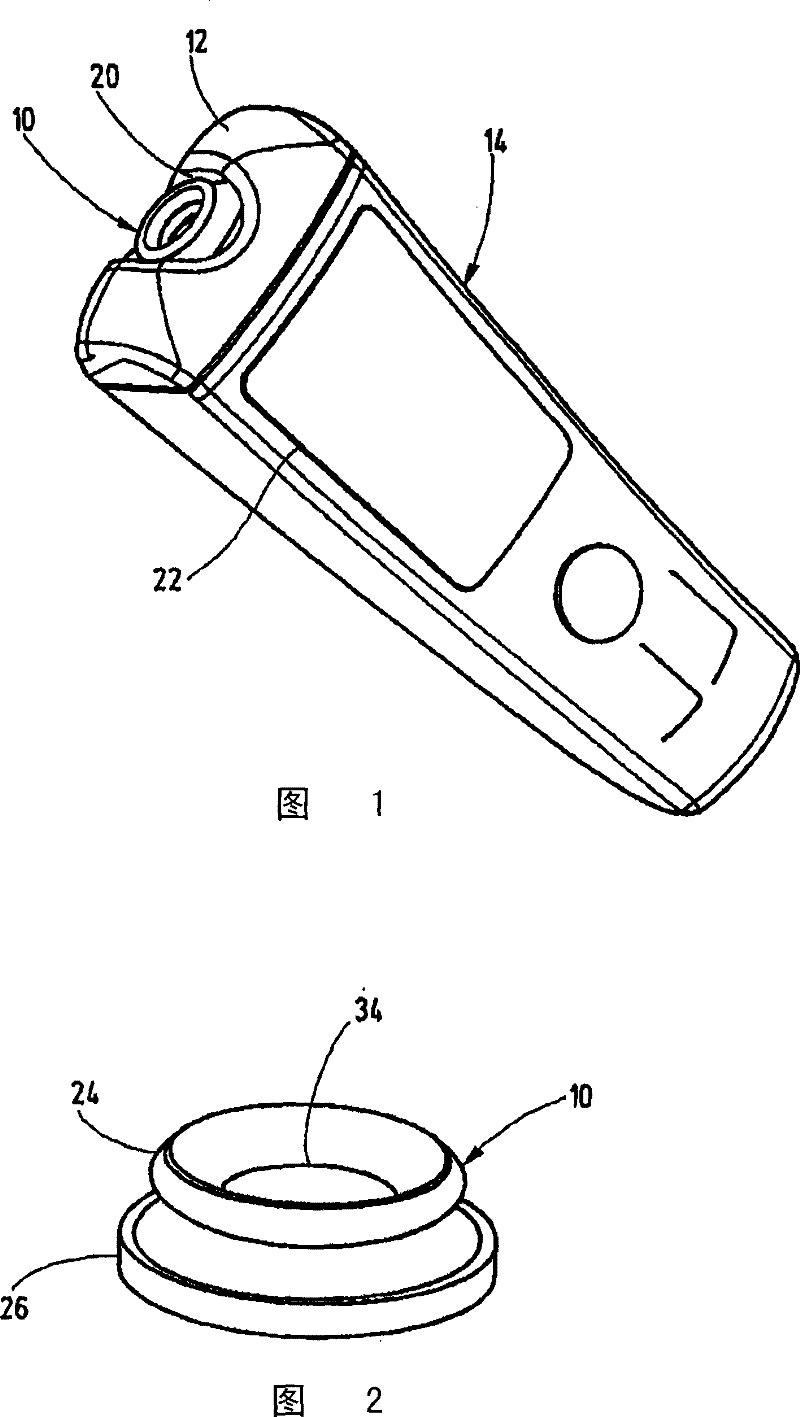
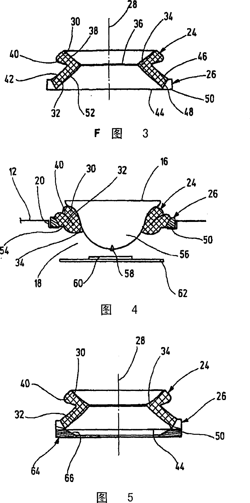
[0041] In order to position the tester's finger pad 16 inside the instrument 18 for blood collection and analysis, the positioning device 10 shown in the drawing can be inserted into the housing 12 of the portable blood glucose meter 14 as an easily replaceable insert.

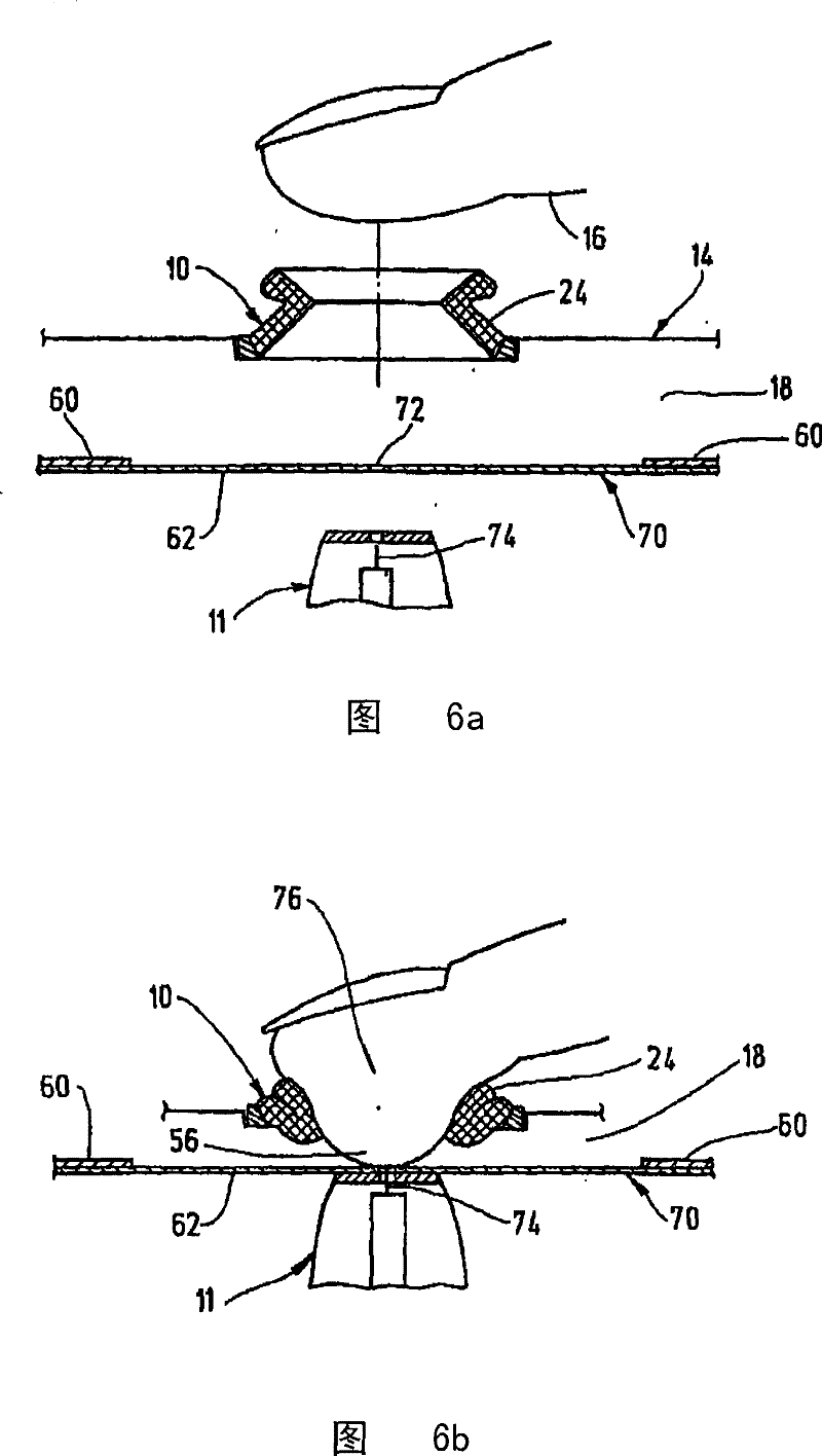
[0042] For this purpose, figure 1 The hand-held instrument 14 shown in FIG. 2 has a fastening point 20 for an exchangeable positioning device 10 on one end face. The device comprises, as an integrated system, inside the device a piercing mechanism 11 for piercing the finger pad and an analysis device 13 for determining the sugar content of the blood collected at the piercing site ( FIG. 6 ). The test result can be displayed to the user through the display unit 22 .

[0043] exist figure 2 and 3As shown most clearly in , the positioning device 10 has a flexible compression element 24 for compressing the finger pad 16 and a dimensionally stable support 26 for fixing the compression element 24 . The compres...


PUM


Abstract

The invention relates to a device and a method for positioning a body part (16) in an analytical device (14) for body fluids, comprising a flexible compression element (24) for placing the body part (16) against it under increasing pressure, and a rigid support part (26) retaining the compression element (24) on a housing (12) of the analytical device (14). The invention is characterized in that the compression element (24) comprises an engaging zone (34) for the body part (16), which can be pushed into the housing (12), so that a withdrawal zone (56) of the body part (16) is exposed in the interior (18) of the housing (12) for the withdrawal of the fluid.

Description

technical field [0001] The invention relates to a device and a method for positioning a body part, in particular a finger pad, on a body fluid analyzer and a corresponding analyzer according to the preambles of the independent claims. Background technique [0002] DE 10026172 A1 discloses a system for extracting body fluids, in which a deformable biconical body acts as a compression unit for increasing the internal pressure in the region of the compressed body part. During this process, the pressing force with a component transverse to the main direction is partly converted into a movement in the secondary direction until the two cone regions, the upper and the lower, rest against each other. In this state, the stenosis that binds the body part is located above the plate-shaped support, while the puncture or blood collection site is still located inside the folded double cone. This requires punctate interventions for the blood collection, at which point there is a risk of ...

Claims


Application Information

Patent Timeline
Patent Type & Authority Patents(China)
IPC IPC(8): A61B5/15
CPCA61B5/151A61B5/157A61B5/14532A61B5/1411A61B5/150068A61B5/150022A61B5/150358A61B5/150412A61B5/150755A61B5/150748
Inventor F·德克
Owner F HOFFMANN LA ROCHE & CO AG
Learn More