Human brain glioma marker glial fibrillary acidic protein (GFAP) autoantibody and applications thereof
A technology of human glioma and autoantibodies, which is applied in the fields of biotechnology and medicine, can solve the problems of high cost of imaging diagnosis technology and the inability to be widely used
- Summary
- Abstract
- Description
- Claims
- Application Information
AI Technical Summary
Problems solved by technology
Method used
Image

Examples
Embodiment 1
[0031] Example 1: Serum collection and its clinical information
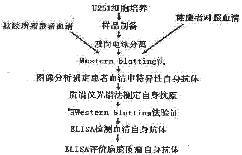
[0032] Serum samples of glioma patients of different grades were collected from Shanghai Huashan Hospital, and 41 healthy people of corresponding age and sex from Huashan Hospital were used as normal controls. All samples were stored in a -80°C freezer. Patient serum samples were classified into astrocytoma, anaplastic astrocytoma, and glioblastoma multiforme according to their respective histopathological analysis. Using proteomics methods, twenty glioblastoma multiforme patient serum samples and their corresponding normal human serum samples were used to detect and screen for autoantibodies. 81 serum samples were used to detect and confirm the presence of GFAP autoantibodies (this research has been approved by the Ethics Committee of Huashan Hospital and Biomedical Research Institute affiliated to Fudan University).
Embodiment 2
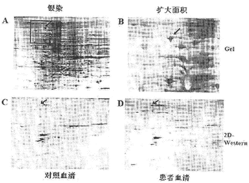
[0033] Example 2: 2D-Western analysis
[0034]U251 brains collected by centrifugation were lysed using urea buffer (8M urea, 2.5M thiourea, 65mM DTT, 4% (w / v) CHAPS, 0.5% (v / v) Biolytes pH 3-10, protease inhibitor cocktail) Glioma cells were quantified using RC DC Protein Assay Kit (Bio-Rad), and 30 micrograms of protein were diluted in rehydration buffer (7M urea, 2M thiourea, 50mM DTT, 4% (w / v) CHAPS, 0.2 % (v / v) Biolytes pH 3-10), and loaded in IPG strips. Isoelectric focusing was performed using Protean IEF Cell (BioRad), followed by SDS-PAGE electrophoresis. Separated proteins were visualized by silver staining or transferred to PVDF membrane overnight with a 100 mA current. The same protein was run on two gels in parallel, one with patient serum for western blotting, and the other for western blotting with normal human serum. After blocking for two hours in TBST containing 4% BSA, the two membranes were incubated in 1:200 dilution of patient and corresponding normal h...
Embodiment 3
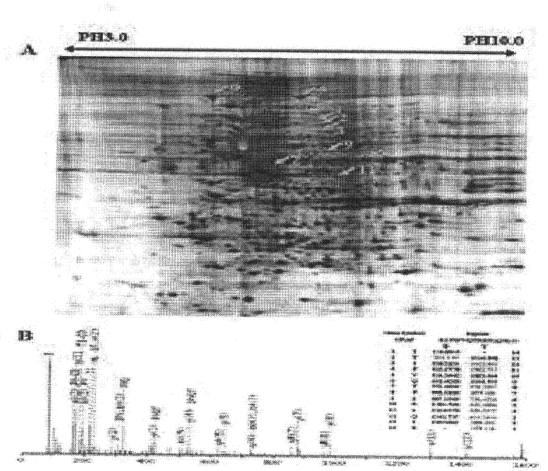
[0035] Embodiment 3: image analysis and protein identification
[0036] LAS-3000 and supporting software Gauge V3.0 software (Fuji, Japan) were used to analyze and identify the difference of immune reaction spots on the membrane incubated with patient and corresponding normal human serum. The corresponding position of each protein spot was confirmed by comparing the pictures of silver-stained 2D gel and western blot. Proteins that only appear in human serum were spot-cut, decolorized, enzymatically hydrolyzed and freeze-dried. The process is to destain the excised strips in 50mM NH4HCO3, 50% acetonitrile, then add 10mM DTT (Amersco, Solon, Ohio) at 56°C for reduction, and incubate in the dark by adding 55mM iodoacetamide (Bio-Rad) Alkylation. The gel pieces were dehydrated with acetonitrile and air dried. 25 mM ammonium bicarbonate containing 0.01 μg / ul trypsin (Promega) was added to carry out in-gel enzymatic hydrolysis at 37 degrees for 16 hours, and then the peptide was ...
PUM
 Abstract
Description
Claims
Application Information
 - R&D Engineer
- R&D Manager
- IP Professional
- Industry Leading Data Capabilities
- Powerful AI technology
- Patent DNA Extraction
Browse by: Latest US Patents, China's latest patents, Technical Efficacy Thesaurus, Application Domain, Technology Topic.
© 2024 PatSnap. All rights reserved.Legal|Privacy policy|Modern Slavery Act Transparency Statement|Sitemap