
Method and device for processing image data obtained by medical equipment

A technology of image data and medical equipment, which is applied in the field of image data processing and can solve problems such as large image color deviations

Active Publication Date: 2016-08-17
EDAN INSTR
View PDF11 Cites 0 Cited by
  • Summary
  • Abstract
  • Description
  • Claims
  • Application Information

AI Technical Summary

Problems solved by technology

[0003] The technical problem to be solved by the present invention is to provide a method for compensating the color temperature of the image data so that the color deviation of the image data is relatively small in view of the above-mentioned defects in the prior art that the color temperature of the image changes due to the difference in the color temperature of the light source. Method and device for processing image data acquired by medical equipment

Method used


Image

  • Method and device for processing image data obtained by medical equipment
  • Method and device for processing image data obtained by medical equipment
  • Method and device for processing image data obtained by medical equipment

Examples


Embodiment Construction

[0042] The embodiments of the present invention will be further described below in conjunction with the drawings.

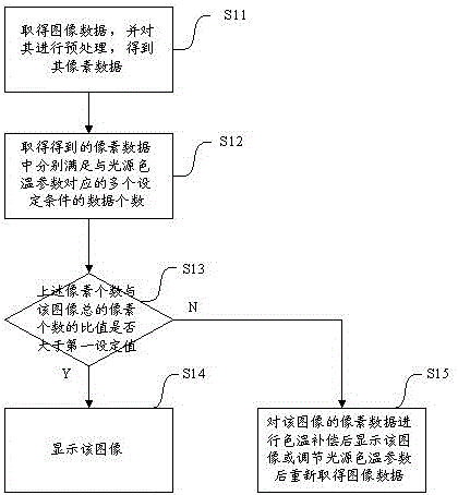
[0043] Such as figure 1 As shown, in the embodiment of the processing method and device for obtaining image data by medical equipment of the present invention, the processing method for obtaining image data includes the following steps:

[0044] Step S11: Obtain image data and preprocess it to obtain its pixel data: In this step, the medical device obtains image data and preprocesses it to facilitate subsequent data processing. In this embodiment, the processing method of obtaining an image by an electronic colposcopy is taken as an example to specifically describe the processing method of obtaining an image from it. As we all know, the existing medical equipment usually obtains images through the CCD image photoelectric sensor. The image signals obtained are electrical signals in the form of analog signals. Before obtaining image data, it is usually necessary to set ...


PUM


Abstract

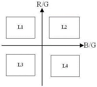
The invention relates to a method for processing image data obtained by a medical device. The method includes the following steps: obtaining the image data, and preprocessing the image data to enable the image data to be stored in a digital pixel data mode; by calculating B / G values and R / G values in the pixel data, judging whether pixels are located in multiple set areas, judging whether a ratio of the number of the pixels located in the multiple set areas to the total number of the pixels is larger than a first set value or not, and if the ratio is not larger than the first set value, carrying out color temperature compensation or correction on the pixel data; if the ratio is larger than the first set value, displaying an image formed by the pixels; determining a color temperature value of the data in one set area to be an image color temperature value, correcting color temperature values of the data in the other set areas to be the color temperature value or correcting a color temperature value of a light source of the medical device, and obtaining the image data again. The invention further relates to a device for achieving the method. The method and device for processing the image data obtained by the medical device have the advantage that the image with the small color deviation is obtained.

Description

Technical field [0001] The present invention relates to image data processing, and more specifically, to a processing method and device for obtaining image data by medical equipment. Background technique [0002] In some medical equipment, it is usually necessary to obtain the patient’s part of the image and display it in a certain form, such as on the display screen, to provide a basis or reference for the operator’s judgment. Electronic colposcopy is one of the more typical equipment. . Generally speaking, electronic colposcopy is commonly used in the diagnosis of lower genital tract diseases and the diagnosis of early cervical cancer. The colposcopy magnifies the images of the cervix, vagina and vulva by a certain multiple, and observes the changes of the epithelium and blood vessels that are invisible to the naked eye under the illumination of the light source. By observing these changes, electronic colposcopy can find cervical erosion, cervical polyps, cervical intraepithe...

Claims


Application Information

Patent Timeline
Patent Type & Authority Patents(China)
IPC IPC(8): H04N9/04H04N9/73H04N5/235
Inventor 章鸿杨天明
Owner EDAN INSTR
Learn More