Automatic positioning of standard planes for real-time fetal heart assessment

A fetal heart, image plane technology, applied in the field of medical diagnostic ultrasound systems, can solve the problems of insufficient volume frame rate and unutilized

Active Publication Date: 2018-02-23
KONINKLJIJKE PHILIPS NV
View PDF12 Cites 0 Cited by
  • Summary
  • Abstract
  • Description
  • Claims
  • Application Information

AI Technical Summary

Problems solved by technology

Unfortunately, in most cases, current volumetric frame rates are not high enough to provide sufficient both spatial and temporal resolution (volume ratio) to make live 3D imaging practical for fetal heart imaging
Furthermore, since only specific 2D image planes are required for fetal heart diagnosis, not much 3D information is utilized

Method used


Image

  • Automatic positioning of standard planes for real-time fetal heart assessment
  • Automatic positioning of standard planes for real-time fetal heart assessment
  • Automatic positioning of standard planes for real-time fetal heart assessment

Examples


Embodiment Construction

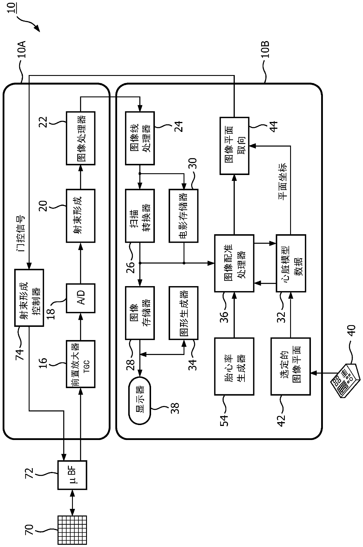
[0014] first reference figure 1 , shows in block diagram form an ultrasound system 10 constructed in accordance with the principles of the present invention. The ultrasound system is configured by two subsystems: a front-end acquisition subsystem 10A and a display subsystem 10B. The ultrasound probe is coupled to an acquisition subsystem comprising a two-dimensional matrix array transducer 70 and a microbeamformer 72 . The microbeamformer contains circuitry that controls the signals applied to the groups of elements ("modules") of the array transducer 70 and performs preliminary processing of the echo signals received by the elements of each group. Microbeamforming in the probe advantageously reduces the number of conductors in the cable between the probe and the ultrasound system, and is described in US Patent 5,997,479 (Savord et al.) and in US Patent 6,436,048 (Pesque).

[0015] The probe is coupled to the acquisition subsystem 10A of the ultrasound system. The acquisiti...


PUM


Abstract

An ultrasound system and method for acquiring standard views of the fetal heart concurrently with real-time imaging are described. Manipulate the matrix array probe until a first standard view such as a four-chamber view is acquired. The first standard view image is matched to a corresponding plane of the first standard view image in a fetal heart model. From the matched planes of the heart model, the orientation of the other standard views is known from the geometrical relationship of the structures within the heart model. This orientation information is used to control the matrix array probe to automatically scan all of the standard view planes simultaneously in real time.

Description

technical field [0001] The present invention relates to medical diagnostic ultrasound systems, and in particular to ultrasound systems capable of performing fetal heart diagnosis by real-time imaging of diagnostically useful image planes. Background technique [0002] Ultrasound imaging is routinely used during pregnancy to evaluate the development of the fetus in the mother's uterus. Ultrasound screening of the fetal heart aims to detect structural abnormalities (typically congenital heart defects or CHDs) and includes analysis of standard two-dimensional (2D) image views of the fetal heart. Typical standard views include a four-chamber view and a view that enables evaluation of the left ventricular (LV) outflow tract and the right ventricular (RV) outflow tract. Other views that may be requested include the five-chamber, three-vessel, and tracheal views. In fact, these views often reveal most of the CHD. The traditional way for a clinician to acquire images of each requ...

Claims


Application Information

Patent Timeline
Patent Type & Authority Patents(China)
IPC IPC(8): A61B8/14A61B8/08A61B8/00
CPCA61B8/0866A61B8/145A61B8/483A61B8/54G06T19/00G06T2210/41G06T2219/028F04C2270/0421G06T7/30G06T15/08G06T17/00G06T2207/10132G06T2207/30048
Inventor J·R·杰戈A·道A·科莱比永L·K·庞弗里
Owner KONINKLJIJKE PHILIPS NV
Why Eureka
Try Eureka