
A serum-containing oral mucosal epithelial cell culture solution
An oral mucosal epithelial and cell culture technology, applied in the field of biomedicine, can solve the problems of increasing cell passages, declining stem cell characteristics, slow growth of oral mucosal epithelial cells, etc., to avoid slow cell growth, achieve large-scale expansion, Guarantees the effect of high-density proliferation
- Summary
- Abstract
- Description
- Claims
- Application Information
AI Technical Summary
Problems solved by technology
Method used
Image

Examples
Embodiment 1
[0024] An embodiment of the present invention provides a serum-containing oral mucosal epithelial cell culture solution, including basal culture solution, serum, essential supplementary factors, cell growth factors, adhesion factors, and regulatory factors for cell proliferation and differentiation.
[0025] Among them, the serum is fetal bovine serum with a concentration of 0.5%-5%, the essential supplementary factors include insulin and transferrin with a concentration of 5-15μg / ml, and the cell growth factors include epidermal growth factor with a concentration of 5-20ng / ml EGF, insulin growth factor IGF-I, transforming growth factor TGFβ and BPE, the adherence factor is fibronectin with a concentration of 5-20ng / ml, and the regulatory factor of cell proliferation and differentiation is retinoic acid and glycerol with a concentration of 1-5μM. Alkyd, Rock Inhibitor Y27632 and CaCl2.
[0026] It should be noted that the basal culture fluid is K-SFM culture fluid or MCDB153 c...
Embodiment 2
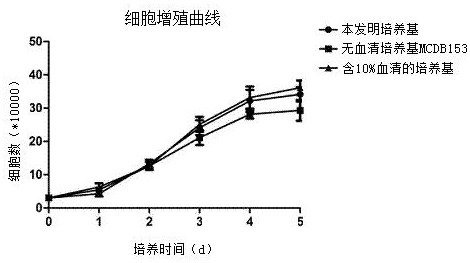
[0049] Using serum-free medium MCDB153, a culture system containing 10% serum and the serum-containing culture solution provided by the embodiment of the present invention to cultivate oral mucosal epithelial cells, a comparative study of the proliferation curve of P6 generation oral mucosal epithelial cells:
[0050] Experimental operation method:
[0051] The P6 oral mucosal epithelial cells in good growth condition were taken. When the oral mucosal epithelial cells were 80%-90% confluent, the cells were washed with PBS, digested with trypsin at 37°C for about 2 min, and the digestion was terminated, and then evenly divided into two Centrifuge in a centrifuge tube. Resuspend the cells using the culture medium provided in the examples of the present invention, the culture system containing 10% serum and the serum-free medium MCDB153, and perform cell counting, inoculate the 24-well plate according to the amount of 3E4 cells, and then place the well plate in Culture in a 5% C...
Embodiment 3
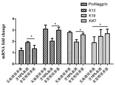
[0054] Serum-free medium MCDB153, F12 / DMEM (1:3) medium system containing 10% fetal bovine serum and the culture medium provided in the examples of the present invention were used to culture primary oral mucosal epithelial cells, and the changes in the content of proteins related to cell proliferation and differentiation Comparative Studies: Experimental How-to:
[0055] Under sterile conditions, the oral mucosa of Kunming mice 1 day after birth was excised, the submucosal tissue was trimmed and cut into tissue pieces about 1 mm × 1 mm in size, and washed 3 to 4 times with PBS containing double antibodies. Put the specimen into 4 mg / ml Dispase II, digest at 37°C for about 15 minutes, and then separate the epithelial layer. After gently washing the epithelial layer with PBS, put it in 0.25% trypsin, digest it at 37°C for about 10 minutes, stop the digestion, pipette into a single cell suspension, centrifuge to discard the supernatant, and inoculate in three different culture me...
PUM
| Property | Measurement | Unit |
|---|---|---|
| concentration | aaaaa | aaaaa |
| concentration | aaaaa | aaaaa |
| concentration | aaaaa | aaaaa |
Abstract
Description
Claims
Application Information