Reaction membrane strip for detecting autoimmune diabetes, preparation method and using method

An autoimmune and reactive membrane technology, applied in the field of biomedicine, can solve problems such as β-cell autoimmune damage, and achieve the effect of improving the detection efficiency and the accuracy of result judgment, and the result judgment is intuitive and accurate

Pending Publication Date: 2017-12-08
SHENZHEN YHLO BIOTECH +1
View PDF5 Cites 4 Cited by
  • Summary
  • Abstract
  • Description
  • Claims
  • Application Information

AI Technical Summary

Problems solved by technology

At present, it is believed that positive ICA indicates the autoimmune da

Method used


Image

  • Reaction membrane strip for detecting autoimmune diabetes, preparation method and using method
  • Reaction membrane strip for detecting autoimmune diabetes, preparation method and using method

Examples


Embodiment 1

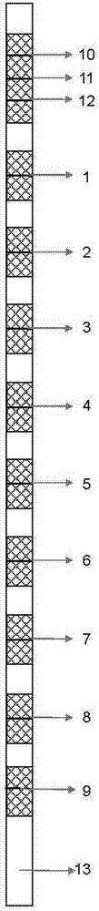
[0090] Please refer to figure 1 and figure 2 , a reaction film strip for autoimmune diabetes detection, the reaction film strip includes a backing plate (13), and more than one detection band and indicator band arranged on the backing plate (13) , the detection bands, the indicator bands, and the detection bands and the indicator bands are parallel to each other;

[0091] The detection zone includes

[0092] the first detection zone (1) coated with GAD antigen,

[0093] A second test strip (2) coated with the IA-2A antigen,

[0094] A third test strip (3) coated with IAA antigen,

[0095] A fourth detection zone (4) coated with ICA antigen,

[0096] The fifth detection zone (5) coated with ZnT-8A antigen,

[0097] A sixth detection zone (6) coated with CP-HA antigen,

[0098] A seventh detection zone (7) coated with SO-XA antigen,

[0099] An eighth detection zone (8) coated with UBE2L3 antigen,

[0100] A ninth detection zone (9) coated with EEF1A1 antigen;

[0101...

Embodiment 2

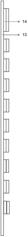
[0109] In the film sticking method, the carrier of the reaction film strip is a double-sided adhesive PVC board (14), and the front side is formed with detection strips (1, 2, 3, 4, 5, 6, 7, 8, 9) and indicator strips (10, 11, 12) said nitrocellulose or nylon film is fixedly bonded, and a transparent PET backing plate (13) is fixedly bonded on the back. The first high-concentration indicating band (10), the second medium-concentration indicating band (11), and the third low-concentration indicating band (12) are drawn by human IgG, or mouse IgG or anti-goat IgG. The enzyme conjugate is horseradish peroxidase-labeled mouse anti-human IgG; the chromogenic substrate is tetramethylbenzidine; the washing solution is 20×Tirs buffer; the sample diluent Tirs buffer containing blocking agent; the stop solution is 0.1 M / L sulfuric acid. The reaction membrane strip in the streaking method directly uses nitrocellulose or nylon membrane 15 as the antigen carrier.

[0110] In order to mak...

Embodiment 3

[0112]GAD, IA-2A, IAA, ICA, ZnT-8A, CP-HA, SO-XA, UBE2L3, and EEF1A1 antigens were tested at a concentration of 0.05-0.1 mg / mL, and human IgG, anti-mouse IgG, or anti-goat IgG were used for quality control According to the first high concentration indicator band (10) with a concentration range of 30-50 μg / mL, the second medium concentration indicator band (11) with a concentration range of 12-25 μg / mL, and a concentration range of 1-10 μg / mL The third low concentration indicator strip (12) is prepared into a corresponding coating concentration for use. According to the size of the immunoblotting spotter, cut the nitrocellulose membrane or nylon membrane into a membrane piece, and coat the above-mentioned configured spotting solution on the membrane piece in a certain order by means of physical adsorption and covalent bonding , forming an independent detection line and judgment indicator band. Put the coated film in a 37°C constant temperature oven to dry for 2 hours, take it ...


PUM


Abstract

The invention provides a reaction membrane strip for detecting autoimmune diabetes, a preparation method and a using method, and belongs to the field of biomedical science. Aiming at the lack of an immunoblotting product special for various immune diabetes experimental examinations and physical examination at present and some defects of an existing immunoblotting agent, the invention provides the reaction membrane strip for detecting the autoimmune diabetes. The reaction membrane strip comprises a bottom lining plate, not less than one detecting zone and not less than one indicating zone, wherein the detecting zones and the indicating zones are arranged on the bottom lining plate; the detecting zones, the indicating zones as well as the detecting zones and the indicating zones are parallel to each other. Compared with the prior art, the reaction membrane strip provided by the invention has the following advantages that various autoantibodies can be detected through one clinic sample and once detection reaction, insufficient sensitivity and specificity of single autoantibody detection and operation complexity of independently detecting autoantibodies related to various diseases one by one are overcome, and the detecting efficiency and the result judgment accuracy are greatly increased.

Description

technical field [0001] The invention belongs to the field of biomedicine, and in particular relates to a reaction membrane strip for autoimmune diabetes detection, a preparation method and a use method. Background technique [0002] The main function of the human immune system is to identify and remove antigenic foreign bodies. Under normal circumstances, the body generally does not produce an immune response to its own tissue components, which is called immune tolerance. Once this tolerance is broken, autoimmune disease occurs. Autoimmune diseases are characterized by the formation of various autoantibodies in serum and damage to multiple organs throughout the body. Among autoimmune diseases, autoimmune diabetes is more common. [0003] Diabetes is mainly classified according to its etiology. In immune-mediated type 1 diabetes, autoantibodies against pancreatic β cells can be detected in the serum of patients, which is a strong evidence of immune damage. Although in the...

Claims


Application Information

Patent Timeline
IPC IPC(8): G01N33/535
CPCG01N33/535G01N2800/042
Inventor 胡鹍辉王栋凯刘尧林俊郑燕萍
Owner SHENZHEN YHLO BIOTECH
Why Eureka
Try Eureka