Probe auxiliary support for ultrasonic examination

A technology of auxiliary bracket and probe bracket, which is applied in ultrasonic/sonic/infrasonic diagnosis, sonic diagnosis, infrasonic diagnosis, etc. It can solve the problems that medical staff cannot accurately control the ultrasonic probe, medical staff's wrist is tired and injured, and the inspection time is prolonged. To achieve the effect of convenient control, convenient cardiac ultrasound diagnosis, and ease of control difficulty

Inactive Publication Date: 2020-05-19
李双晖
View PDF0 Cites 2 Cited by
  • Summary
  • Abstract
  • Description
  • Claims
  • Application Information

AI Technical Summary

Problems solved by technology

[0002] As is known, cardiac ultrasound examination mainly includes several types such as m-mode ultrasound and Doppler. Usually, several methods are carried out at the same time during the examination. Based on the comprehensive analysis of the results, it is possible to determine the size of the heart structure, whether the valves are abnormal, or It can judge whether there is congenital heart disease or valvular disease according to the condition of blood flow. In addition, according to the situation of cardiac ejection, it can judge whether there is cardiac insufficiency. It can also judge cardiomyopathy and wall changes after myocardial infarction. As a result, when performing an echocardiographic examination on a patient, medical personnel need to hold an ultrasonic probe and attach it to the upper part of the patient's chest for examination. The examination time will be extended according to the patient's condition, and sometimes it will be extended to one hour. It is a long examination process In the process, because the medical staff's wrists are suspended in the air and there is no focus, it not only causes fatigue and injury to the medical staff's wrists, but also causes the medical staff to be unable to accurately control the ultrasonic probe, which makes the ultrasonic probe press on the upper part of the patient's chest for a long time, causing local pain to the patient.

Method used


Image

  • Probe auxiliary support for ultrasonic examination
  • Probe auxiliary support for ultrasonic examination
  • Probe auxiliary support for ultrasonic examination

Examples


Embodiment Construction

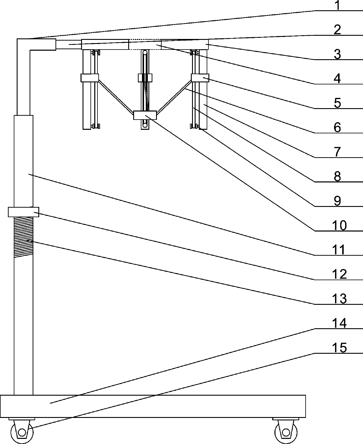
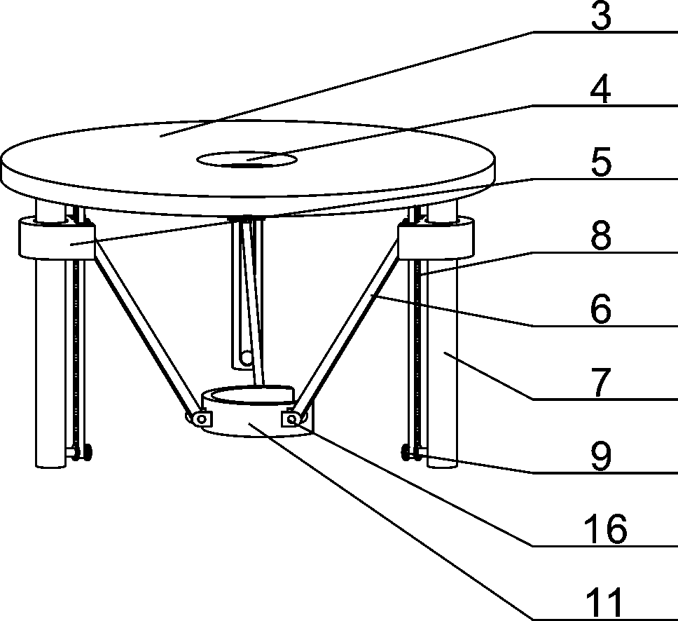
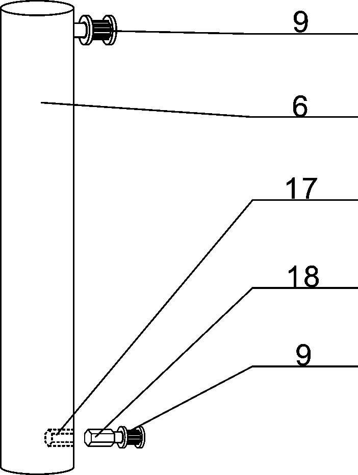
[0030] The present invention is described in further detail now in conjunction with accompanying drawing. These drawings are all simplified schematic diagrams, which only illustrate the basic structure of the present invention in a schematic manner, so they only show the configurations related to the present invention.

[0031] combined with Figure 1~6In the given auxiliary probe bracket for ultrasonic inspection, the sliding bracket is composed of a bottom plate 14, a support rod 11 and an "L"-shaped rod 1, and a support rod 11 is provided on one side of the surface of the bottom plate 14. The upper surface of the rod 11 is provided with an external thread 13, and an elbow support 13 threaded with the external thread 13 is provided on the pole 11, and the elbow support 12 is supported by a collar 28 and plate 30, an insertion ear 32 is provided on one side of the collar 28, and an inserting plate 31 matching the insertion ear 32 is provided on the side of the support plate ...


PUM


Abstract

The invention discloses a probe auxiliary support for ultrasonic examination and relates to a medical auxiliary device. A support rod is arranged on one side of the surface of a bottom plate, an L-shaped rod in telescopic fit with the support rod is arranged at the upper end of the support rod, an ultrasonic probe support is fixedly connected to the other end of the L-shaped rod, a top plate is fixedly connected to the other end of the L-shaped rod, an opening is formed in the middle of the surface of the top plate, three vertical rods distributed in a isosceles triangle shape are arranged onthe lower surface of the top plate, sliding blocks in sliding fit with the vertical rods are arranged on the three vertical rods respectively, connecting rods are movably connected to inner side surfaces of the three sliding blocks, and the other ends of the three connecting rods are movably connected with the outer wall of a C-shaped clamping part. Lifting adjustment of an ultrasonic probe can becontrolled at multiple angles, and limitation and fixation of the height of the ultrasonic probe can be realized, so that the ultrasonic probe can be accurately and slightly attached to the surface of the chest of a patient.

Description

technical field [0001] The invention relates to a medical auxiliary device, in particular to a probe auxiliary bracket for ultrasonic examination. Background technique [0002] As is known, cardiac ultrasound examination mainly includes several types such as m-mode ultrasound and Doppler. Usually, several methods are carried out at the same time during the examination. Based on the comprehensive analysis of the results, it is possible to determine the size of the heart structure, whether the valves are abnormal, or It can judge whether there is congenital heart disease or valvular disease according to the condition of blood flow. In addition, according to the situation of cardiac ejection, it can judge whether there is cardiac insufficiency. It can also judge cardiomyopathy and wall changes after myocardial infarction. As a result, when performing an echocardiographic examination on a patient, medical personnel need to hold an ultrasonic probe and attach it to the upper part...

Claims


Application Information

Patent Timeline
Patent Type & Authority Applications(China)
IPC IPC(8): A61B8/00
CPCA61B8/4218A61B8/4405A61B8/4411A61B8/4444
Inventor 李双晖
Owner 李双晖
Why Eureka
Try Eureka