A Segmentation Method of Left and Right Ear Regions Based on CT Scanning Images of Temporal Bone

A region segmentation and CT image technology, applied in the field of medical image processing, to achieve the effect of fast operation, simple and effective method, and avoidance of interference

Active Publication Date: 2022-03-18
FUDAN UNIV
View PDF4 Cites 0 Cited by
  • Summary
  • Abstract
  • Description
  • Claims
  • Application Information

AI Technical Summary

Problems solved by technology

[0004] So far, there is no special algorithm for the segmentation and extraction of left and right ear regions from temporal bone CT images

Method used


Image

  • A Segmentation Method of Left and Right Ear Regions Based on CT Scanning Images of Temporal Bone
  • A Segmentation Method of Left and Right Ear Regions Based on CT Scanning Images of Temporal Bone
  • A Segmentation Method of Left and Right Ear Regions Based on CT Scanning Images of Temporal Bone

Examples


Embodiment Construction

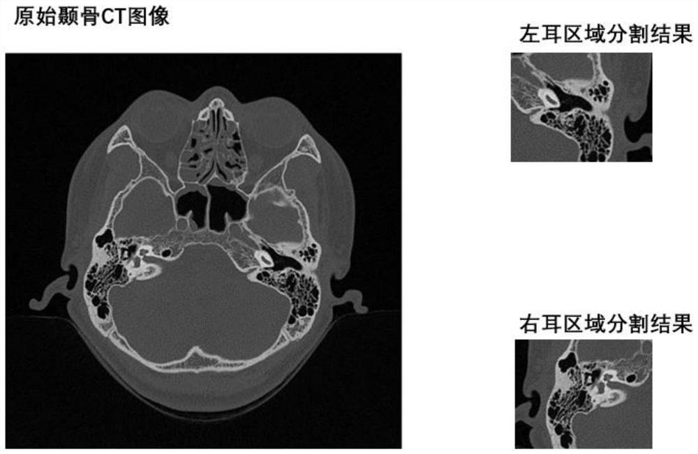
[0029] For an input temporal bone CT picture I, the left and right ear regions are segmented and extracted. The specific steps are:

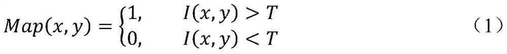
[0030] (1) Use the threshold T=500 to perform effective area binarization on I to obtain a binarized feature map. Calculate the upper boundary UP, lower boundary DOWN, left boundary LEFT, and right boundary RIGHT of the effective area according to the feature map. Crop I according to the boundary, and use the Bicubic algorithm to interpolate the CT image of the effective area after clipping to enlarge it to 512×512;

[0031] (2) Cropping of left and right ear regions and discarding of irrelevant regions. The range of the right ear area is: the upper boundary up takes 200, the lower boundary down takes 300, and the left boundary left r Take 1, the right boundary right r Take 250; the range of the left ear area is: the upper boundary up takes 200, the lower boundary down takes 300, and the left boundary left l Take 263, the right boundary rig...


PUM


Abstract

The invention belongs to the technical field of medical image processing, in particular to a method for segmenting left and right ear regions based on temporal bone CT plain scan images. This algorithm is used to segment the left and right ear regions in the temporal bone CT image of the head, so that the computer can perform subsequent processing on the ear regions intensively. The present invention comprises the following steps in sequence: removing the invalid area of ​​the head CT image; cutting out the left and right ear areas in the image according to the proportion; respectively arranging and storing the images of the left and right ear areas. Experimental results show that, for plain-scanned temporal bone CT images, this method can remove invalid black regions, discard information of other irrelevant parts, and accurately extract left and right ear regions.

Description

technical field [0001] The invention belongs to the technical field of medical image processing, in particular to a method for segmenting left and right ear regions based on temporal bone CT plain scan images. Background technique [0002] With the development of computer science, intelligent medical care has become a major technological innovation to improve the level of modern medical care. Its advantages in various aspects are getting more and more recognition and attention. Among them, medical image processing technology is one of the key parts to promote the development of intelligent medical care, and it is also a very important application link of intelligent medical care. [0003] In the application scenario of CT imaging diagnosis in otology, doctors can only obtain CT images of the entire temporal bone at this stage, so as to diagnose the patient's ear lesions. However, when doctors actually make a diagnosis, they only observe and base on the ear area in the CT i...

Claims


Application Information

Patent Timeline
Patent Type & Authority Patents(China)
IPC IPC(8): G06T7/11G06T7/136
CPCG06T7/11G06T7/136G06T2207/10081G06T2207/20132G06T2207/30008
Inventor 王云峰颜波李健李吉春谭伟敏
Owner FUDAN UNIV
Why Eureka
Try Eureka