Light source emission detection probe with integrated structure

A technology for detecting probes and integrated structures, which is applied in the medical field and can solve problems such as troublesome interconnection and integration, inconvenient disassembly and replacement, etc.

Inactive Publication Date: 2021-09-14
SECOND AFFILIATED HOSPITAL OF COLLEGE OF MEDICINEOF XIAN JIAOTONG UNIV
View PDF0 Cites 0 Cited by
  • Summary
  • Abstract
  • Description
  • Claims
  • Application Information

AI Technical Summary

Problems solved by technology

In terms of signal synchronization, photoelectric marking is used to achieve high-precision signal synchronization triggering, but the interconnection and integration of multiple probes is very troublesome, and it is not easy to disassemble and replace

Method used


Image

  • Light source emission detection probe with integrated structure
  • Light source emission detection probe with integrated structure
  • Light source emission detection probe with integrated structure

Examples


Embodiment Construction

[0027] In order to make the technical means, creative features, goals and effects achieved by the present invention easy to understand, the present invention will be further described below in conjunction with specific embodiments.

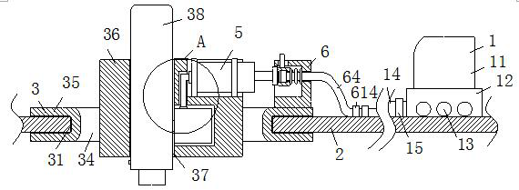
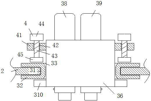
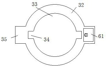
[0028] Such as Figure 1-Figure 7As shown, a light source emission detection probe with an integrated structure according to the present invention includes a light source emission assembly 1, the light source emission assembly 1 includes a light source emitter 11, and the light source emitter 11 is fixedly connected through a mounting base 12 On the surface of the headgear 2, the side surface of the installation base 12 is provided with several female optical fiber interfaces 13, and the insides of the several female optical fiber interfaces 13 are inserted with female optical fiber main bodies 14, and the female optical fiber main bodies 14 pass through the first A fixed buckle 15 is fixedly connected to the surface of the headgear 2, the upper s...


PUM


Abstract

The invention relates to the field of medical treatment, and particularly relates to a light source emission detection probe with an integrated structure. The light source emission detection probe comprises a light source emission assembly, wherein the light source emission assembly comprises a light source emitter; the light source emitter is fixedly connected to the surface of a head sleeve through a mounting base; a plurality of female optical fiber interfaces are formed in the side surface of the mounting base; female optical fiber main bodies are inserted into the plurality of female optical fiber interfaces; the female optical fiber main bodies are fixedly connected to the surface of the head sleeve through first fixing buckles; and a plurality of mounting mechanisms are arranged on the upper surface of the head sleeve. A light source receiving assembly and a connecting mechanism are arranged and are used for automatically butting the interfaces when a cylindrical block is fixed, so that stable transmission of signals is facilitated, and the installation is convenient; after the cylindrical block is inserted into a circular groove and rotates, the cylindrical block is driven by a pressing mechanism to ascend, and meanwhile, a receiving probe ascends; a sleeve is pushed by a bevel ring to move back to compress a spring; and after the cylindrical block is fixed, the sleeve and the bevel ring are completely closed and butted.

Description

technical field [0001] The invention relates to the medical field, in particular to a light source emission detection probe with an integrated structure. Background technique [0002] No matter whether the active cells or tissues of the human body are in the static state or the active state, they will produce regular electrical phenomena closely related to the life state, which is called bioelectricity. Through high-precision weak bioelectrical signal acquisition technology, various electrophysiological signals of the human body can be collected, including EEG, EMG, ECG, etc., and the obtained data can be analyzed and stored. Among them, EEG is the weakest of all bioelectrical signals and the most difficult signal to collect. It contains a large number of electrophysiological activity information of brain nerve cells, which can reflect information such as people's thinking activities and body movements. Electroencephalogram (EEG) as a It is a relatively mature brain functio...

Claims


Application Information

Patent Timeline
Patent Type & Authority Applications(China)
IPC IPC(8): A61B5/369A61B5/00A61B5/386A61B5/291A61B5/256
CPCA61B5/369A61B5/386A61B5/291A61B5/256A61B5/0075A61B5/0042A61B5/6803
Inventor 李立博惠艳娉
Owner SECOND AFFILIATED HOSPITAL OF COLLEGE OF MEDICINEOF XIAN JIAOTONG UNIV
Why Eureka
Try Eureka