Automatic glandular and tubule detection in histological grading of breast cancer

a breast cancer and histological grading technology, applied in the field of histopathology, can solve the problems of time-consuming and subjective process, adversely affecting the estimation of tubule percentage, and not ideal

Active Publication Date: 2019-10-15
VENTANA MEDICAL SYST INC
View PDF5 Cites 1 Cited by
  • Summary
  • Abstract
  • Description
  • Claims
  • Application Information

AI Technical Summary

Problems solved by technology

Tubule score is traditionally calculated by manually estimating the percentage of glandular regions in the tumor that form tubules, which is a time-consuming and subjective process.
Since these methods only associate the lumen and its closest nuclei, they cannot handle cases in which a lumen is surrounded by multiple layers of nuclei, which can adversely affect the estimation of tubule percentage.
Additionally, these methods rely on analysis of images that represent only a small segment of the whole slide tissue, which is not ideal.
To our knowledge, no current methods are available that can address both of these deficiencies.

Method used


Image

  • Automatic glandular and tubule detection in histological grading of breast cancer
  • Automatic glandular and tubule detection in histological grading of breast cancer
  • Automatic glandular and tubule detection in histological grading of breast cancer

Examples


Embodiment Construction

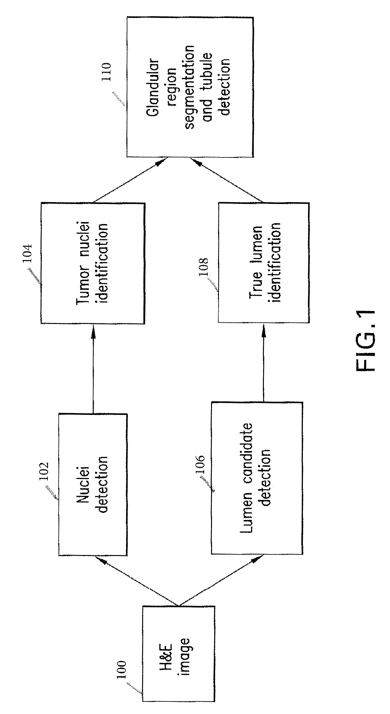
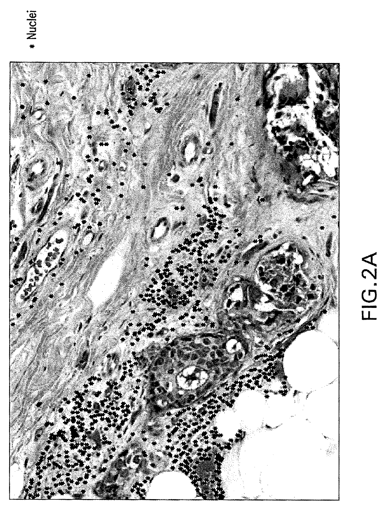
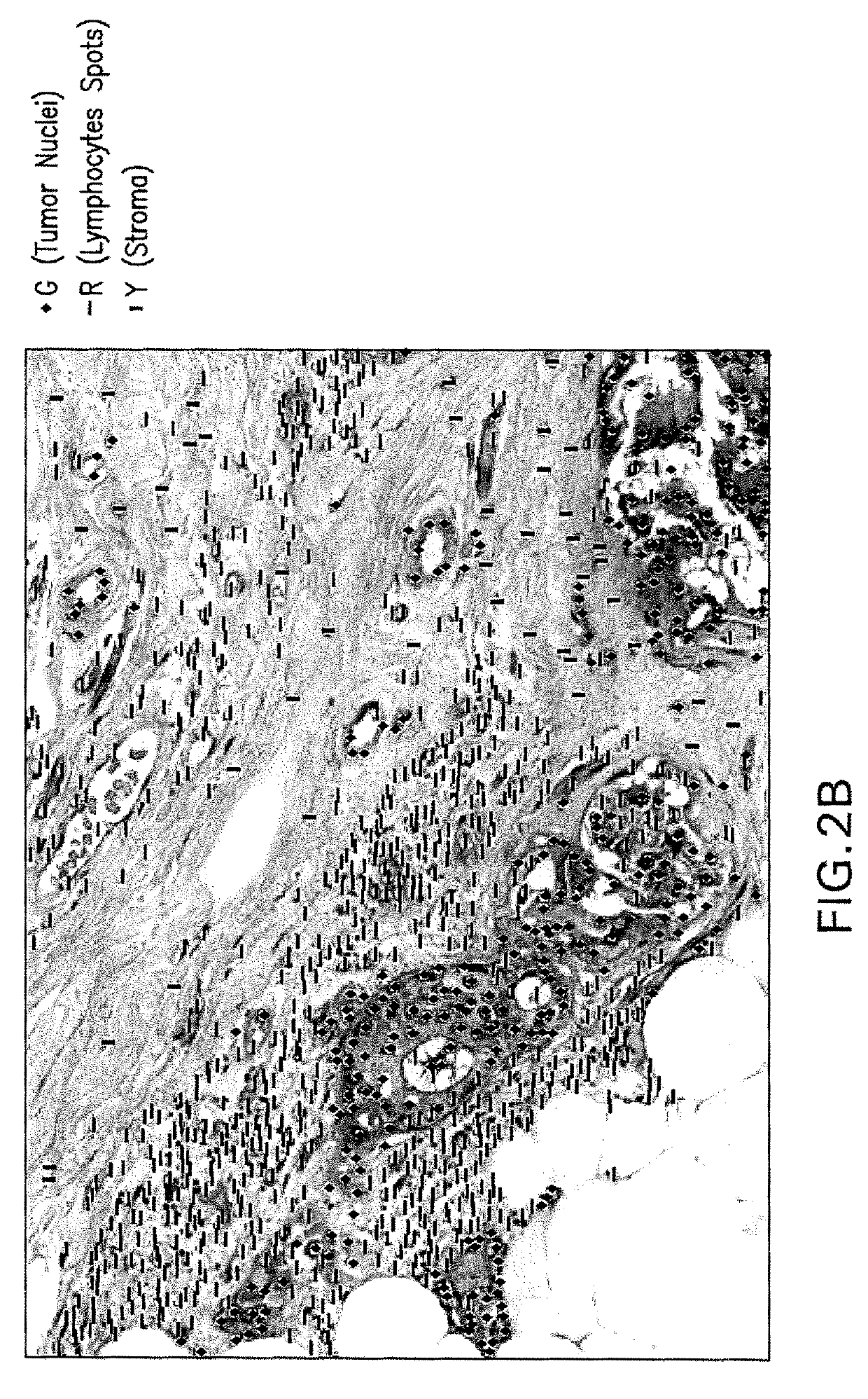
[0016]Methods, systems, and apparatuses are provided for automatically identifying tubule glandular regions and non-tubule glandular regions in an image of a breast tissue sample by: (a) identifying tumor nuclei and true lumina in the image; and (b) identifying glandular regions by grouping the tumor nuclei with neighboring lumina and neighboring tumor nuclei, wherein glandular regions containing true lumina are classified as tubule glandular regions, and wherein glandular regions lacking true lumina are classified as non-tubule glandular regions. Tubule percentage and tubule score can then be calculated. Tubule percentage (TP) as understood herein is the ratio of the tubule area to the total glandular area. The analysis can be applied to whole slide images and can resolve tubule areas with multiple layers of nuclei. These methods, systems, and apparatuses are particularly useful in pathological scoring systems for breast tumors, such as NHS. Other utilities and advantages of the pr...


PUM


Abstract

Methods, systems, and apparatuses for automatically identifying glandular regions and tubule regions in a breast tissue sample are provided. An image of breast tissue is analyzed to detect nuclei and lumen candidates, identify tumor nuclei and true lumen from the candidates, and group tumor nuclei with neighboring tumor nuclei and lumina to define tubule glandular regions and non-tubule glandular regions of the image. Learnt supervised classifiers, such as random forest classifiers, can be applied to identify and classify the tumor nuclei and true lumina. Graph-cut methods can be applied to group the tumor nuclei and lumina and to define the tubule glandular regions and non-tubule glandular regions. The analysis can be applied to whole slide images and can resolve tubule areas with multiple layers of nuclei.

Description

REFERENCE TO RELATED APPLICATIONS[0001]This patent application is a continuation of International Patent Application No. PCT / EP2015 / 067007 filed Jul. 24, 2015, which claims the benefit of U.S. Provisional Application No. 62 / 030,009, filed Jul. 28, 2014. Each of the above patent applications is incorporated herein by reference as set forth in its entirety.BACKGROUND OF THE DISCLOSURE[0002]The present disclosure relates to the use of image analysis in the field of histology to identify glandular and tubule glandular regions in breast cancer tissue.[0003]Histological grading is an important step in breast cancer prognosis. In the popular Nottingham Histologic Score (NHS) system for breast cancer grading (c.f. Breast Cancer Research and Treatment, 1992, Volume 22, Issue 3, pp 207-219, The Nottingham prognostic index in primary breast cancer, Marcus H. Galea et. al.), the pathologist analyzes tissue for tubule formation, nuclear pleomorphism and mitotic activity in the tumor regions and ...

Claims


Application Information

Patent Timeline
Patent Type & Authority Patents(United States)
IPC IPC(8): G06K9/62G06T7/00G06K9/66G06T7/40G06K9/00G06T7/11G06T7/13G06K9/46
CPCG06V20/69G06V20/695G06V20/698G06V10/7635G06F18/24323G06F18/285G06F18/214G06T7/11G06T7/13G06T7/0012G06T7/40G06T2207/30024G06T2207/30068G06T2207/30096
Inventor BARNES, MICHAELCHEFD'HOTEL, CHRISTOPHECHUKKA, SRINIVASNGUYEN, KIEN
Owner VENTANA MEDICAL SYST INC
Why Eureka
Try Eureka