
Device for separation of corneal epithelium

a technology of corneal epithelium and device, which is applied in the field of ocular surgical devices, can solve the problems of increasing the cost of the device, increasing the incidence of device failure, and not being fully satisfactory to many practitioners, and achieves the effects of simple and elegant design and construction, simple and elegant assembly sequence, and easy manipulation

Inactive Publication Date: 2005-03-10
SIGHTRATE
View PDF99 Cites 20 Cited by
  • Summary
  • Abstract
  • Description
  • Claims
  • Application Information

AI Technical Summary

Benefits of technology

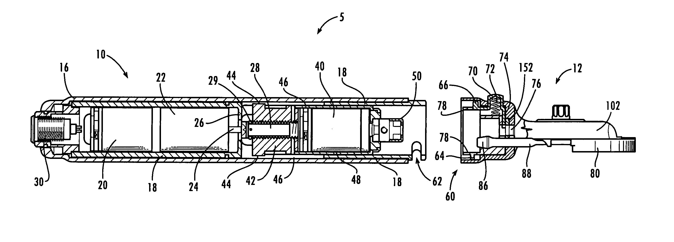
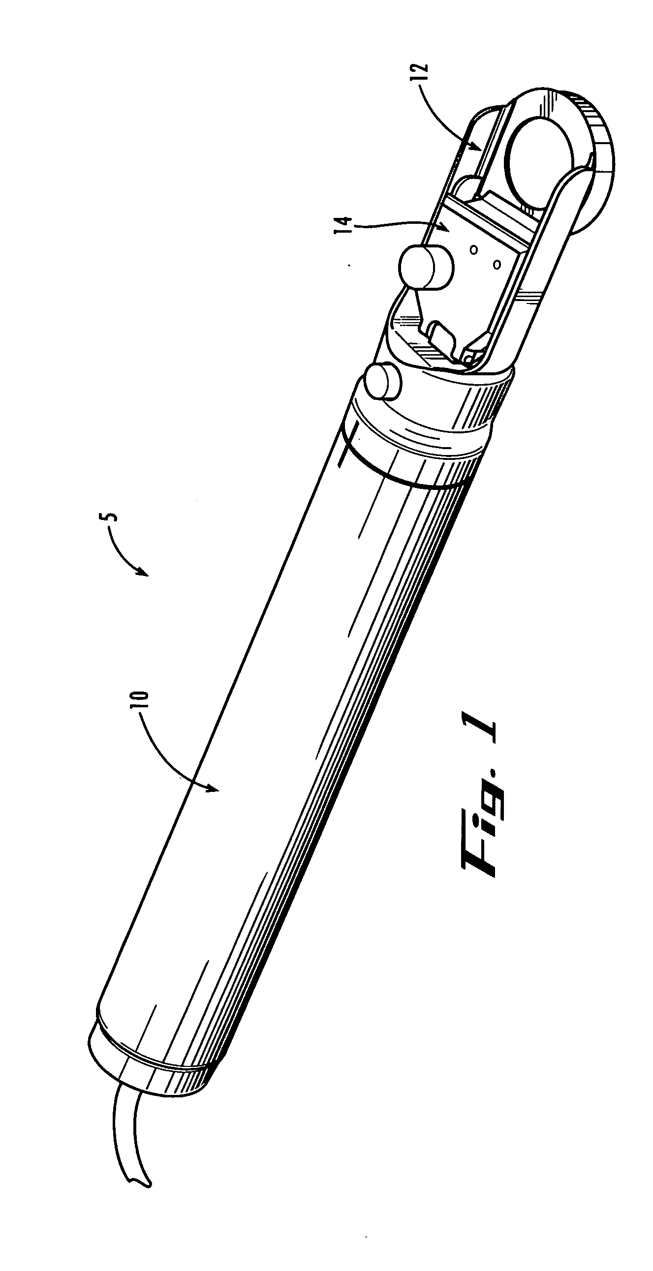
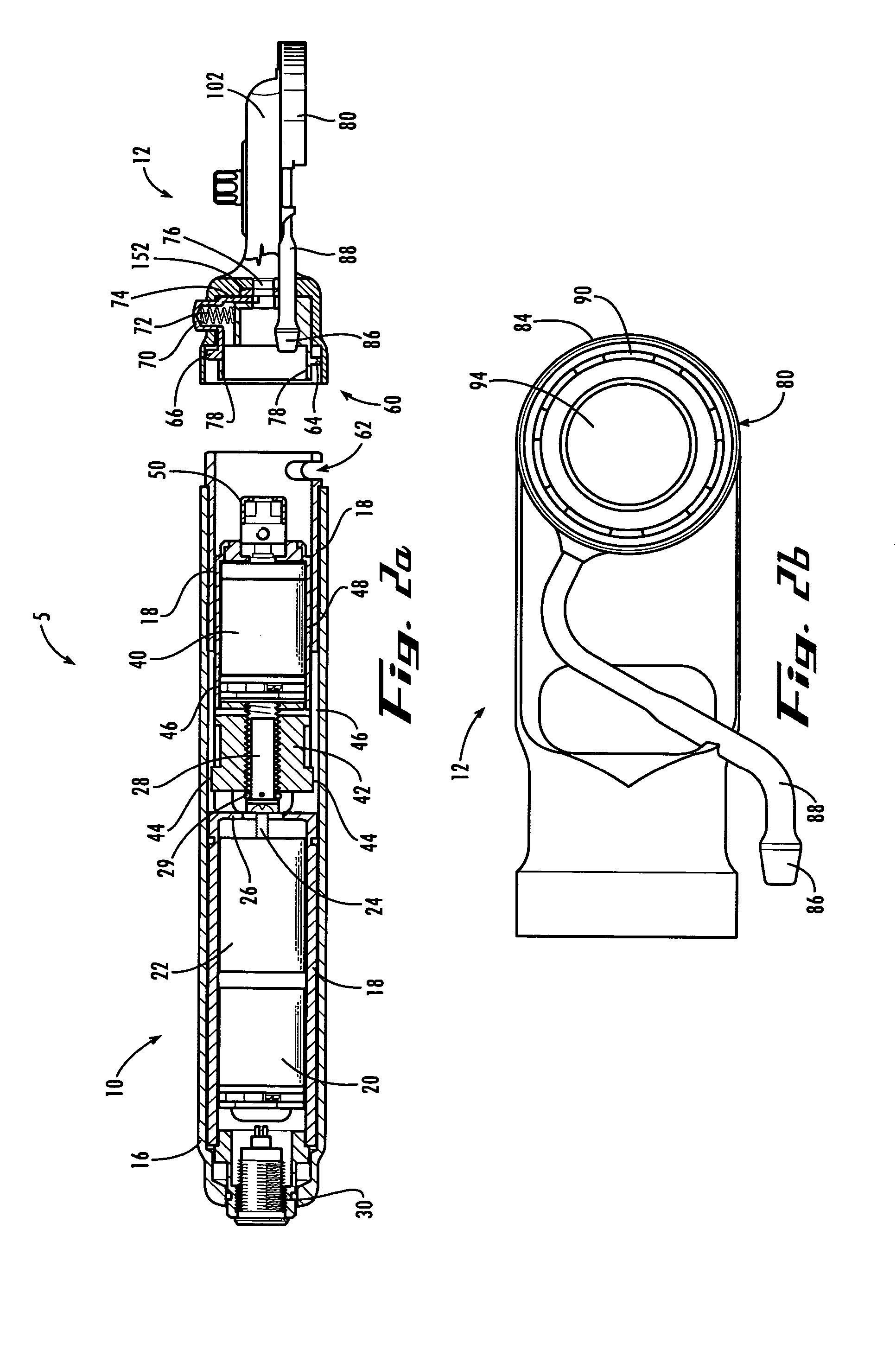
[0007] In example forms, the present invention is an improved drive tool for use in ocular surgery. In preferred applications, the tool is well suited to driving a blunt polymeric separator to separate the corneal epithelium from underlying Bowman's layer for subsequent corneal reshaping. In alternate embodiments, the device of the present invention may find application as a drive tool for standard sharp microkeratome blades, as in traditional LASIK procedures. The device of the present invention is preferably simple and elegant in design and construction, minimizing the necessary components and optimizing their assembly configuration, thereby resulting in a compact, ergonomic and easily manipulated surgical tool. In particularly preferred embodiments, the device is configured for comfortable one-hand operation by the practitioner. The device preferably also includes integral assembly interlocks, simplifying the proper assembly sequence and preventing improper assembly and disassembly, and preventing operation if the device is not fully and correctly assembled. The device is preferably configured for connection and use with standard suction and drive controllers that many practitioners will already have available, and with which practitioners are familiar and experienced in operating.

Problems solved by technology

Previously known microkeratome devices have not proven fully satisfactory to many practitioners.
For example, it has been found that many known microkeratome devices are complex and difficult to assemble and disassemble properly, potentially leading to difficulties in sterilization for reuse, interruptions in the surgical procedure, unduly adding to the cost of the devices, and increasing the incidence of device failure.
It has also been found that many known microkeratome devices are bulky and unwieldy in use, potentially resulting in user fatigue and increasing the risk of errors during a procedure.
Previously known microkeratome devices also have not been found to be well suited to the newly developed procedures for separation of corneal epithelium.

Method used


Image

  • Device for separation of corneal epithelium
  • Device for separation of corneal epithelium
  • Device for separation of corneal epithelium

Examples


Embodiment Construction

[0027] The present invention may be understood more readily by reference to the following detailed description of the invention taken in connection with the accompanying drawing figures, which form a part of this disclosure. It is to be understood that this invention is not limited to the specific devices, methods, conditions or parameters described and / or shown herein, and that the terminology used herein is for the purpose of describing particular embodiments by way of example only and is not intended to be limiting of the claimed invention. Also, as used in the specification including the appended claims, the singular forms “a,”“an,” and “the” include the plural, and reference to a particular numerical value includes at least that particular value, unless the context clearly dictates otherwise. Ranges may be expressed herein as from “about” or “approximately” one particular value and / or to “about” or “approximately” another particular value. When such a range is expressed, anothe...


PUM


Abstract

A drive tool for optical surgery includes a handpiece containing a traverse motor and an oscillating motor, wherein the oscillating motor is axially driven through the handpiece by the traverse motor. A head assembly including a suction ring is mounted to the handpiece, and a separator drive assembly is positioned therein for advancement imparted by the traverse motor and oscillation imparted by the oscillating motor.

Description

CROSS-REFERENCE TO RELATED APPLICATION [0001] This application claims priority benefit to U.S. Provisional Patent Application Ser. No. 60 / 500,874, filed Sep. 5, 2003, the entirety of which is hereby incorporated by reference herein.TECHNICAL FIELD [0002] The present invention relates generally to ocular surgical devices, and more particularly to a surgical apparatus and method for separating the epithelium layer of a cornea from the underlying Bowman's layer with minimal trauma to the epithelium and Bowman's layer. BACKGROUND OF THE INVENTION [0003] Microkeratome devices are widely used in LASIK (Laser-Assisted In Situ Keratomilousis) procedures. LASIK permanently changes the shape of the cornea, the clear covering of the front of the eye, using an excimer laser. A microkeratome is used to cut a corneal flap, typically containing an overlying layer of corneal epithelium, Bowman's layer, and a portion of the stroma by slicing through the stroma, dividing it into at least two distinct...

Claims


Application Information

Patent Timeline
IPC IPC(8): A61F9/013
CPCA61F9/013A61F9/007
Inventor WOODS, STEPHEN P.
Owner SIGHTRATE
Learn More